Advanced Medical Diagnostics
The Advanced Medical Diagnostics and Lab-on-a-Chip research theme builds upon over 20 years of research in Glasgow, and has a strong track record in many aspects of bioengineering, especially those associated with advanced biomedical diagnostics, biosensors, cell engineering, Lab-on-a-Chip and Bionanotechnology.
Our work is supported through strong national and international collaborations with academia and industry, particularly in the development of biophotonics and acoustic manipulation of fluids for point-of-care diagnostics, optical and optoelectronic tools for the manipulation and characterisation of particles, microfluidic devices for proteomics, the development of cell based technologies for the pharmaceutical industry.
Research topics
- Advanced diagnostics
- LOC for developing world diagnostics
- Micromanipulation for advanced diagnostics
- Microrheology
- Single cell analysis
- Bio nano photonics
Staff
| Professor Jon Cooper | Professor Thomas Franke |
| Dr Andrew Glidle | Profesor Alasdair Clark |
| Dr. Chunxiao Hu | Dr Julien Reboud |
| Dr Manlio Tassieri | Professor Huabing Yin |
| Dr. Morteza Amjadi | Dr. Jiabao Xu |
Advanced Diagnostics
Pr. Jon Cooper, Dr. Alasdair Clark, Dr. Andrew Glidle, Dr. Julien Reboud, Pr. Huabing Yin, Dr. Chunxiao Hu, Dr. Jiabao Xu.
In the field of medical sensing, the need to capture diagnostic information in remote settings using low power disposable systems will be key.
Microtechnologies present opportunities for creating advanced diagnostic tools, which are faster, more sensitive, and use less analyte than their traditional macro scaled counterparts. “Lab-on-a-Chip” techniques are promising improved information at reduced costs.
This in turn enables to perform diagnosis or testing away from the centralised laboratory, providing analysis of samples in remote settings (as might be the case for in the field environmental monitoring) or for medical diagnostics (as would be appropriate either for point-of-care sensing or for implantable in vivo sensing).
To achieve this, even in the simplest cases, a Lab-on-a-Chip needs to carry out a number of discrete operations that may include sample manipulation, fluid or reagent movement and sensing. Research into Lab-On-a-Chip (LOC) technologies looks into how different processes can be brought onto one microfabricated chip.
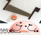
We have several projects that use LOC techniques to create new diagnostic devices described in the information on researchers associated with this theme. This includes projects using acoustics (surface acoustic waves) to manipulate fluids and the particles (or pathogens) within them to create a tool-box of integrated diagnostic functions; the development of ultrasensitive biosensors based on photonics and acoustics ; and optoelectronic devices to manipulate rare cell populations.
LOC for developing world diagnostics
Pr. Jon Cooper, Dr. Alasdair Clark, Dr. Andrew Glidle, Dr. Steven Neale, Dr. Julien Reboud, Pr. Huabing Yin
The developing world presents unique challenges for diagnosing diseases. Tests must be inexpensive and capable of being carried out in areas without reliable infrastructure but with the same requirements for sensitivity and specificity as traditional lab based diagnostic methods. Lab-on-a-Chip technologies offer a way to combat these problems by miniaturising standard methods producing quicker and cheaper results, or by producing new diagnostic tools with no macroscopic equivalent.
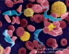
Parasitic infections continue to exert a disproportionate burden on the world's poorest people. Hundreds of millions of people are infected with parasites that cause diseases like malaria and the trypanosomiasis (HAT), a disease of sub-Saharan Africa. The twenty-first century has seen significant progress in reducing the incidence of the disease. However, the drugs used can be dangerous and should only be given to patients positively identified as harbouring parasites. This can be exceedingly difficult since in many cases there are fewer than 100 parasites/ml of blood, amongst billions of red blood cells. If the World Health Organisation's declared intention to eliminate HAT by 2030 is to be reached, enabling diagnosis in individuals with sub-clinical levels of infection will be important.
We are working with our collaborators in the Wellcome Trust Centre for Molecular Parasitology to create new diagnostic tools using lab-on-a-chip technologies including microfluidics, optical tweezers, dielectrophoresis, Optoelectronic Tweezers (OET) and acoustic tweezing.
Micromanipulation for Advanced Medical Diagnostics
Dr. Steven Neale, , Pr. Thomas Franke, Dr. Alasdair Clark, Dr. Andrew Glidle, Dr. Manlio Tassieri, Pr. Jon Cooper
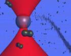
By placing forces onto microscopic biological samples, information can be gathered about the properties of cells or fluids in healthy and diseased states, which translate into information on the health of the patient. Forces can be used to stretch or indent cells, which leads to details on their elasticity, or they can be used to differentiate between species of cells, helping to detect the presence of pathogens or parasites.
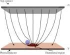
Micromanipulation methods currently being investigated include optical tweezers, optoelectronic tweezers, acoustic manipulation and dielectrophoresis. Each of these techniques has different strengths and limitations making them more or less suitable for particular applications, such as the diagnosis of Human African Trypanosomaisis (HAT also know as sleeping sickness) and using the Young's Modulus of erythrocytes as a biomarker for aging and obesity.
Microrheology
Dr. Manlio Tassieri, Pr. Jon Cooper,
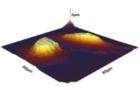
Microrheology is a branch of rheology, which is usually concerned with the studies of the flow of liquids. Microrheology shares the same principles as those of conventional bulk rheology, but works on micron length scales and microliter volumes. It is concerned with the linear viscoelastic properties of materials, such as the viscosity of a fluid.
Microrheological tools are considered essential for biophysical studies, because they match the range of sensitivity required to study a wide range of biological processes. With the exception of Atomic Force Microscopy (AFM), the techniques involved are all based on the observation of either the free or driven motion of micron-sized beads suspended in the solution under investigation. The time-dependent beads trajectories are directly related to the viscoelastic properties of the solution.
The insights gained from Microrheology studies are of great interest to a wide scientific community, including molecular and cell biologists, biochemists, microbiologists, geneticists, and medical researchers.
Single Cell Analysis
Pr. Huabing Yin, Pr. Jon Cooper, Dr. Andrew Glidle, Dr. Massimo Vassalli, Pr. Thomas Franke, Dr. Chunxiao Hu, Dr. Jiabao Xu.
Individual cells have subtle differences even in an isogenic population. Much evidence shows that individual heterogeneity is of pivotal importance to cell function and fate. We have developed a broad range of techniques including micro-fabrication, micro-manipulation and micro-optics, allowing single cell study with statistically meaningful data.
Using these techniques, the position and microenvironment of a cell can be controlled with the use of microfluidics, optical tweezing and dielectrophoresis. Cells can be physically probed, as is the case with Atomic Force Microscopy (AFM) for their mechanical properties, or optically probed, as in Raman imaging, providing the intrinsic chemical fingerprint of a cell. This physical and chemical information is emerging as a new class of cellular biomarker and we use this in the investigation of cell differentiation, cancer development and cell phenotypic identification.
Bionanophotonics
Dr. Alasdair Clark, Dr. Andrew Glidle, Pr. Jon Cooper
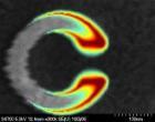
Nanofabrication techniques developed for the fabriaction of electronic components can also be used to create devices with novel biological applications. For example the plasmonic properties of nanostructured metals can be used to significantly improve the sensitivity of biosensing techniques based on the vibrational spectroscopy, Surface Enhanced Raman Scattering (SERS).
One project using this technology aims to use state of the art electron-beam lithography to fabricate arrays of novel, crescent shaped, nano-split-ring resonators (see figure) that can be used as sensing surfaces for bio-molecular detection. Such structures possess multiple, polarisation dependent, plasmon resonances that can be tuned to desired wavelengths through nanometre control of particle geometry.