

Pixelated STEM
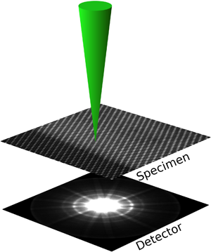 Pixelated STEM is the future of STEM imaging. Traditionally, STEM imaging has been performed mainly with fast integrating detectors, such as annular dark field or bright field detectors. Much work at Glasgow has used split detectors to perform differential phase contrast imaging. But there is much more information in the back focal plane than this, and introducing a fast and noise-free pixelated detector into the microscope allows a huge range of different features of the sample to be imaged using different features of the diffraction pattern. These include, but are not limited to:
- Phase contrast imaging of atoms and nanoscale objects using ptychography
- Imaging of the magnetisation inside materials and thin films using the disc deflection
- Other uses of differential phase contrast, for example to investigate electric fields
- Fluctuation microscopy of medium range order in glasses
- Imaging of the 3D ordering in crystals using high angle scattering into Higher Order Laue Zones
Many of these have been investigated using an EPSRC Research grant "Fast Pixel Detectors: a paradigm shift in STEM imaging" (EP/M009963/1 and EP/M010708/1) led by Dr Ian MacLaren and Dr Damien McGrouther at Glasgow and Prof. Peter D. Nellist at the University of Oxford.
In order to perform this work, a suitable detector is required and much work has gone into integrating a Medipix3 detector into our JEOL ARM200F, as described below.
The results of the pixelated STEM are very large data files and turning these into useful, physically meaningful images is a "big-data" problem. Some details of our work in this area are described below, together with Open-Source software links.
Development of transformational electron detectors for investigation of advanced materials
 Led by Dr Damien McGrouther, a collaboration across the Particle Physics Detectors group (Maneuski & O’Shea) and the Institute for Gravitational Research (Perreur-Lloyd) has developed novel imaging detectors for transmission electron microscopes (TEMs). Harnessing technologies developed at Glasgow and CERN, the ability to unambiguously detect and count individual high energy electrons at MHz rates is enabling the recording of images with greater fidelity, direct filming of dynamically evolving structures and novel scanned mode imaging modalities in TEMs.
Through research stretching back over eight years [1-9], more recently supported by an EPSRC research grant, EP/M009963/1, the potential benefits provided by particle physics technologies, such as the Medipix and Timepix families of hybrid pixel detector have been investigated. Recent successes [1-7] have demonstrated the transformative benefits of these detectors and provided the motivation to develop commercial detectors, enabling widest access to researchers studying advanced materials in academia and industry.
Supported by the University’s Impact Acceleration accounts, EP/K503903/1 & EP/R511705/1, and in commercial collaboration with Quantum Detectors Ltd, the team have developed commercial prototypes and held a scientific demonstration event, away from Glasgow, at the leading ePSIC national facility. The culmination of this phase of development resulted in the agreement with Quantum Detectors of a royalty bearing license agreement, through which designs and know-how are being exploited as part of Quantum Detector’s product offering in the TEM market place [link]. The first commercial detectors resulting from this are being delivered to clients from summer 2017 onwards. This collaboration has enabled entry into a new market for Quantum Detectors and over a three year period it is expected that tens of high-value detector systems will be sold with an approximate value of £1-2M. Commercial success will result in financial and staff growth for the company and continue to support the UK’s reputation as a leading nation in the development and characterisation of advanced materials.
Collaboration between the team and Quantum Detectors Ltd is continuing in two areas. At a fundamental level, through the award of an STFC funded CASE PhD studentship, ST/P002471/1, the “next next” generation of TEM detector technologies are being explored in Glasgow and the STFC facilities at Harwell, Oxfordshire. Commercial development is intended to continue* through a joint project employing a design engineer to develop a larger detector array product and to provide specialist TEM integration support.
* currently being negotiated
Software for Pixelated STEM
fpd
A python package for fast pixelated detector data storage, analysis and visualisation.
pixSTEM
Python library for post processing of pixelated STEM datasets, which includes virtual detectors (BF, ADF), differential phase contrast through center of mass, radial integration, and many convenience functions. Most importantly, it includes functionality for out-of-core processing, meaning at the whole dataset does not have to be loaded into memory. This is especially useful for the pixelated STEM datasets, which can easily be 50 GB+. Builds upon HyperSpy.
fpd_live_imaging
https://fast_pixelated_detectors.gitlab.io/fpd_live_imaging/
Python library for real time processing of pixelated STEM data, allowing for live imaging (1000+ fps) of features while acquiring them during a STEM experiment. Currently it works with the Medipix3 detector, and includes the standard virtual detectors (ADF, BF), and differential phase contrast imaging which enables imaging of magnetic domains.
merlin_interface
https://fast_pixelated_detectors.gitlab.io/merlin_interface/
Python library for interfacing with a Medipix3 detector through the Merlin readout system. Allows for (almost) full automation of acquisition with the Medipix3.
pixelated DPC
https://github.com/matkraj/pixelatedDPC
GPU accelerated analysis of disk image position in pixelated STEM data with subpixel resolution. Specifically for imaging of magnetisation using disk deflection, but also useful in other contexts where disk deflection or distortion reveals useful information.

Publications
 Characterisation of the Medipix3 detector for 60 and 80 keV electrons
J A. Mir, R Clough, R MacInnes, C Gough, R Plackett, I Shipsey, H Sawada, I MacLaren, R. Ballabriga, D Maneuski, V O'Shea, D McGrouther, A I Kirkland
Ultramicroscopy 182 (2017) 44- Electron ptychographic phase imaging of light elements in crystalline materials using Wigner distribution deconvolution
H Yang, I MacLaren, L Jones, G T Martinez, M Simson, M Huth, H Ryll, H Soltau, R Sagawa, Y Kondo, C Ophus, P Ercius, L Jin, A Kovács, P D Nellist
Ultramicroscopy, 180 (2017) 173-179 - Characterisation of amorphous molybdenum silicide (MoSi) superconducting thin films and nanowires,
A Banerjee, L Baker, A Doye, M Nord, R Heath, K Erotokritou, D Bosworth, Z Barber, I MacLaren, R Hadfield
Superconductor Science and Technology, 30 (2017) 084010 - Internal structure of hexagonal skyrmion lattices in cubic helimagnets
D McGrouther, R J Lamb, M Krajnak, S McFadzean, S McVitie, R L Stamps, A O Leonov, A N Bogdanov, Y Togawa
New Journal of Physics, 18 (2016) 095004. - Developing Rapid and Advanced Visualisation of Magnetic Structures Using 2-D Pixelated STEM Detectors
M Nord, M Krajnak, R Bali, G Hlawacek, V Liersch, J Fassbender, S McVitie, G W Paterson, I MacLaren, D McGrouther
Microscopy and Microanalysis, 22 [S3] (2016) 530-531. - Towards Mapping Perovskite Oxide 3-D Structure Using Two-Dimensional Pixelated STEM Detector,
M Nord, A Ross, I Hallsteinsen, T Tybell, I MacLaren
Microscopy and Microanalysis, 22 [S3] (2016) 476-477. - Pixelated detectors and improved efficiency for magnetic imaging in STEM differential phase contrast,
M Krajnak, D McGrouther, D Maneuski, V O' Shea, S McVitie
Ultramicroscopy 165 (2016) 42 - 4D STEM: High efficiency phase contrast imaging using a fast pixelated detector
H Yang, L Jones, H Ryll, M Simson, H Soltau, Y Kondo, R Sagawa, H Banba, I MacLaren, P D Nellist
J. Phys.: Conf. Ser. 644 (2015) 012032 - Medipix2 as a highly flexible scanning/imaging detector for transmission electron microscopy
A MacRaighne, G V Fernandez, D Maneuski, D McGrouther, V O’Shea
Journal of Instrumentation 6, C01047 (2011) - Medipix2/Timepix detector for time resolved Transmission Electron Microscopy
R Beacham, A Mac Raighne, D Maneuski, V O’Shea, S McVitie, D McGrouther
Journal of Instrumentation 6, C12052 (2011)