

Positively selected modifications in the pore of TbAQP2 allow pentamidine to enter Trypanosoma brucei
Published: 14 September 2020
Research by the Institute's Professor Harry De Koning Group has uncovered a new drug resistance mechanism, involving mutations in an aquaporin, by the parasite Trypanosoma brucei, which causes African sleeping sickness.
Research by the Institute's Professor Harry De Koning Group has uncovered a new drug resistance mechanism, involving mutations in an aquaporin, by the parasite Trypanosoma brucei.
African sleeping sickness is a potentially deadly illness caused by the parasite, and although treatable, many of the current treatments are old and becoming increasingly ineffective.
For instance, resistance is growing against pentamidine, a drug used in the early stages in the disease, as well as against melarsoprol, which is deployed when the infection has progressed to the brain.
Usually, cases resistant to pentamidine are also resistant to melarsoprol, but it is still unclear why, as the drugs are chemically unrelated.
Studies have shown that changes in a water channel called aquaglyceroporin 2 (TbAQP2) contribute to drug resistance in African sleeping sickness, suggesting that it plays a role in allowing drugs to kill the parasite.
This molecular ‘drain pipe’ extends through the surface of T. brucei, and should allow only water and glycerol in and out of the cell. In particular, the channel should be too narrow to allow pentamidine or melarsoprol to pass through.
One possibility is that, in T. brucei, the TbAQP2 channel is abnormally wide compared to other members of its family.
Alternatively, pentamidine and melarsoprol may only bind to TbAQP2, and then ‘hitch a ride’ when the protein is taken into the parasite as part of the natural cycle of surface protein replacement.
In this study, published in eLife on 7 August 2020, Professor De Koning aimed to tease out these hypotheses.
Computer models of the structure of the protein were paired with molecular dynamics simulations and engineered changes in the key areas of the channel to show that T. brucei AQP2 provides a much broader gateway into the cell than has been observed for similar proteins in other species.
In addition, genetic analysis showed that this version of TbAQP2 has been actively selected for during the evolution process of T. brucei. This suggests that the parasite somehow benefits from this wider aquaglyceroporin variant.
This is a new resistance mechanism, and it is possible that aquaglyceroporins are also larger than expected in other infectious microbes.
This work by the De Koning group, therefore, provides an invaluable insight into how other germs may become resistant to drugs.
Prof De Koning, Professor of Parasite Biochemistry and Pharmacology, explained: "Antimicrobial resistance is a major concern in the control of infectious disease.
"This work describes a completely new mechanism of drug resistance in the blood parasite that causes African sleeping sickness.
"Furthermore, the discovery has potential ramifications for drug treatments in other pathogens as well."
Positively selected modifications in the pore of TbAQP2 allow pentamidine to enter Trypanosoma brucei
- Ali H Alghamdi, Jane C Munday, Gustavo Daniel Campagnaro, Dominik Gurvic, Fredrik Svensson, Chinyere E Okpara, Arvind Kumar, Juan Quintana, Maria Esther Martin Abril, Patrik Milić, Laura Watson, Daniel Paape, Luca Settimo, Anna Dimitriou, Joanna Wielinska, Graeme Smart, Laura F Anderson, Christopher M Woodley, Siu Pui Ying Kelly, Hasan MS Ibrahim, Fabian Hulpia, Mohammed I Al-Salabi, Anthonius A Eze, Teresa Sprenger, Ibrahim A Teka, Simon Gudin, Simone Weyand, Mark Field, Christophe Dardonville, Richard R Tidwell, Mark Carrington, Paul O'Neill, David W Boykin, Ulrich Zachariae, Harry P De Koning
- eLife 2020;9:e56416 DOI: 10.7554/eLife.56416
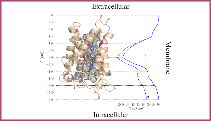
Image modelling (top) by Molecular Dynamics, performed in collaboration by Ulrich Zachariae, University of Dundee.
The figure shows the TbAQP2 protein in light brown, and the pentamidine molecule in dark blue, modelled inside the pore, which is indicated by a mesh. The energy curve to the right indicated the energy of the interaction, showing it is optimally bound when right in the middle of the pore, as predicted by our model.
First published: 14 September 2020