Lightsheet Microscopy
In lightsheet microscopy a fluorescent sample has a sheet of light passed through it from one or two directions. The sample is then imaged perpendicularly to the lightsheet. It is a very quick and gentle process designed for specimens of >100um thickness. With the CVR setup we can image up to 1cm3 with a sample that has been chemically cleared (in essence it’s made transparent).
The CVR’s Zeiss Z1 lightsheet microscope is the only Zeiss Z1 lightsheet microscope installed in Scotland. It provides flexibility in the CVR’s capacity for deep imaging; it can gently and quickly image large specimens, fixed (cleared or aqueous) or live, detailing dynamic biological processes and providing context to higher resolution imaging modalities already located within the CVR such as super-resolution light microscopy and cryo-electron microscopy.
Note for local users: The local user pages provide details of equipment curators and locations.
The CVR’s Zeiss Z1 has the following elements:
- Three lasers of 405nm, 488nm and 561nm to image fluorophores that emit in the blue, green and red areas of the spectrum.
- Double sided illumination (estimated imaging depth from each side is approx. 500um when specimen isn’t cleared. When the specimen is cleared the limitation on imaging depth relates to the effectiveness of the clearing and the size of the specimen that will fit in the specimen chamber. The maximum size of specimen imaged so far is 1cm3.
- Filters for imaging of blue, green and red channels:
- Filter module BP 460-500 (eg DAPI, CFP)
- Filter Module BP 505-545 (eg GFP)
- Filter Module BP 575-615 (eg Cy3)
- Filter Module LP 585 (eg mCherry)
- A light-tight housing with full environmental control.
- A sCMOS PCO.Edge Camera - a fast and sensitive detection system with high signal:noise ratio and high dynamic range.
- Zeiss’s patented method of pivoting the scan head to remove striping/shadowing artefacts.
- Flexible optics for use with aqueous and cleared specimens:
- 5x air objective (NA) for use with samples of 1.33-1.37 refractive index.
- 20x immersion objectives (with zoom optics allowing max mag of 50x) to image:
- cleared specimens of refractive indices 1.42-1.48 (NA=1.0)
- aqueous specimens of RI 1.33-1.37 (NA=1.0)
- Horizontal sample geometry allowing multiple viewing angles and specimen rotation.
- Overview camera to allow accurate positioning of the specimen.
Lightsheet Microscopy
Some views around the CVR’s lightsheet microscope
|
|
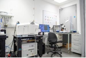
The CVR’s Zeiss Z1 lightsheet microscope. |
|
|
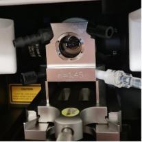
A view of one of the Z1 sample chambers in position for imaging. The specimen is inserted into the imaging space from the top of the microscope and is suspended within the specimen chamber. A laser generated lightsheet is passed through the specimen on the left and right of this chamber and the objective for imaging is located at the back of the chamber. |
|
|
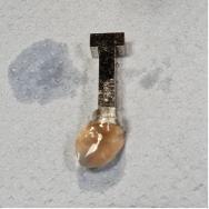
An example of a biological specimen prepared for lightsheet microscopy. The method of mounting the specimen largely depends on its the size and shape. In this example the specimen has been cleared with ethyl cinnamate and is mounted on a metal stub. |
Examples of images acquired using the CVR’s lightsheet microscope
|
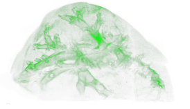
Mosquito malpighian tubules. The image is a snapshot of a multi-image Z stack that was acquired with the Z1 lightsheet and rendered with Imaris software. Lipid within the malpighian tubules is shown in green. |
|
|
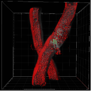
Image of influenza virus-infected mouse lung. This image is a montage of approx. 100 000 separate images (50 fields of view, each of Z stacks with 2000 images) which were acquired with the Z1 lightsheet as individual images and stitched together into a montage using Imaris Stitcher software. Influenza virus is tagged with ZsGreen. |
|