Super-Resolution Microscopy at the CVR
Super-resolution microscopy refers to microscopy that improves resolution beyond the diffraction limitation of light. At the CVR we have two systems capable of super-resolution microscopy.
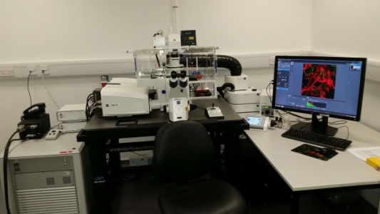
Zeiss LSM 880 Confocal Microscope with Airyscan FAST
The Zeiss LSM 880 utilises Zen 2 (black) software. The system is fitted with a choice of autofocus options and an incubator incorporating full CO2 and temperature control allowing for extended live-cell time-lapse imaging. Other fitted hardware:
|
Objective lenses |
Laser lines |
Detectors |
|
10x (dry) – DIC ready |
405nm (eg DAPI) |
2 x PMTs |
|
20x (dry) – DIC ready |
458nm (eg CFP) |
1 x GaAsP |
|
40x (oil) – DIC ready |
488nm (eg GFP) |
1 x Airyscan |
|
63x (oil) – DIC ready |
514nm (eg YFP) |
|
|
|
561nm (eg Alexa 568) |
|
|
|
594nm (eg Alexa 594) |
|
|
|
633nm (eg Alexa 633) |
|
The quantum efficiency of the GaAsP and Airyscan detectors provides significant improvement in signal:noise. In comparison to the CVR’s LSM 710, which has PMT detectors. The LSM 880 also offers improved scan speed.
Incorporation of an Airyscan detector greatly improves the speed of acquisition but more importantly provides super-resolution capability using the same tried and tested methodology in specimen preparation that is employed in confocal microscopy. The Airyscan enhances theoretical resolution of fluorescent markers to 120nm when combined with modern computational deconvolution methods, comparing favourably with the 240nm xy resolution limitation of the confocal modes and 400nm diffraction-limited resolution anticipated from images acquired using the CVR’s widefield microscopes.
The addition of an Airyscan FAST module to the LSM880 provides further functionality to the microscope. It has increased the speed of Airyscan acquisition 4-fold and provides an expanded range of image resolutions. The associated workstation also allows for synchronised Airyscan processing which further speeds up the image acquisition process.
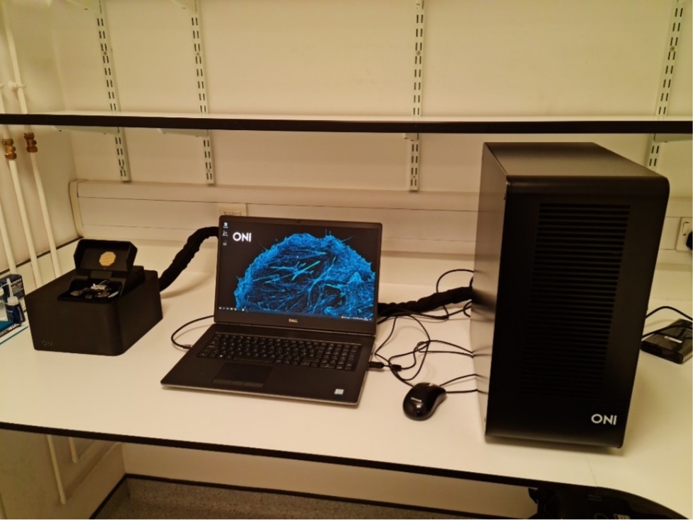
ONI Nanoimager
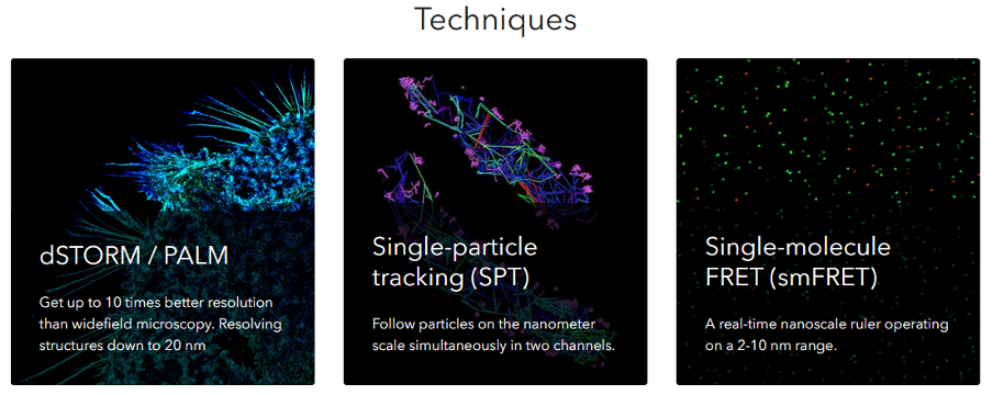
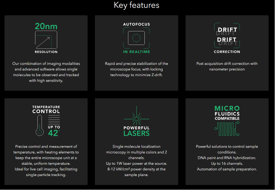
A further step in resolution improvement is provided by our newest optical microscope, the compact ONI Nanoimager with built-in analysis software. This system enables visualization, tracking and imaging of single molecules in living cells. It incorporates multiple super-resolution modes; PALM, dSTORM, single molecule FRET and simultaneous spatio-temporal super-resolution (SPT) and will provide theoretical resolution of 20nm. This has only recently been installed in the CVR (@April 2022) but will be an invaluable tool in elucidating molecular interactions in host and pathogen.
Note for local users: The local user pages provide details of equipment curators and locations.