HIGH CONTENT IMAGING
The CVR has a range of high content imaging systems encompassing both confocal and widefield modalities. Resolution is not the most important factor with any of these instruments, rather each system has improved the CVR’s capability in identifying the interactions and spatial relationships of molecular species and populations in infected specimens.
Note for local users: The local user pages provide details of equipment curators and locations.
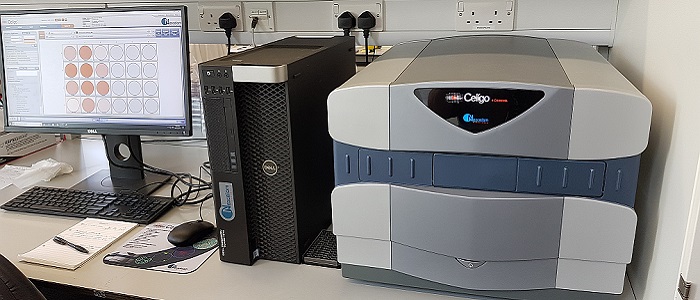
Nexcelom Celigo Imaging Cytometer
The Celigo imaging cytometer is the CVR’s workhorse instrument for imaging adherent and suspension cells in microwell plates. Provided with multiple algorithms to detect, segment and analyse cells across a range of end-point assays the Celigo has recently had a server and satellite workstation upgrade to provide additional data storage and image processing capability, so streamlining workflow. It is a bench-top, micro-well plate based, brightfield and fluorescent imaging system. Its proprietary optics and scanning system enables fast imaging of entire wells while maintaining consistent illumination and contrast out to the well edge, for accurate identification of all cells within each well. Additionally the integrated analysis software allows for segmentation and quantification of suspension or adherence cells within 6, 12, 24, 48, 96, 384 or 1536-well plastic plates.
The LEDs and filters installed are:
|
LEDs |
Excitation |
Beam splitter |
Emission |
Some fluorophores that work |
|
Blue |
377/50 |
409 |
470/22 |
Hoechst, DAPI |
|
Green |
483/32 |
506 |
536/40 |
FITC, GFP, Alexa 488 |
|
Red |
531/40 |
593 |
629/53 |
Texas Red, Alexa 555/568 |
Find out more at Nexcelom.
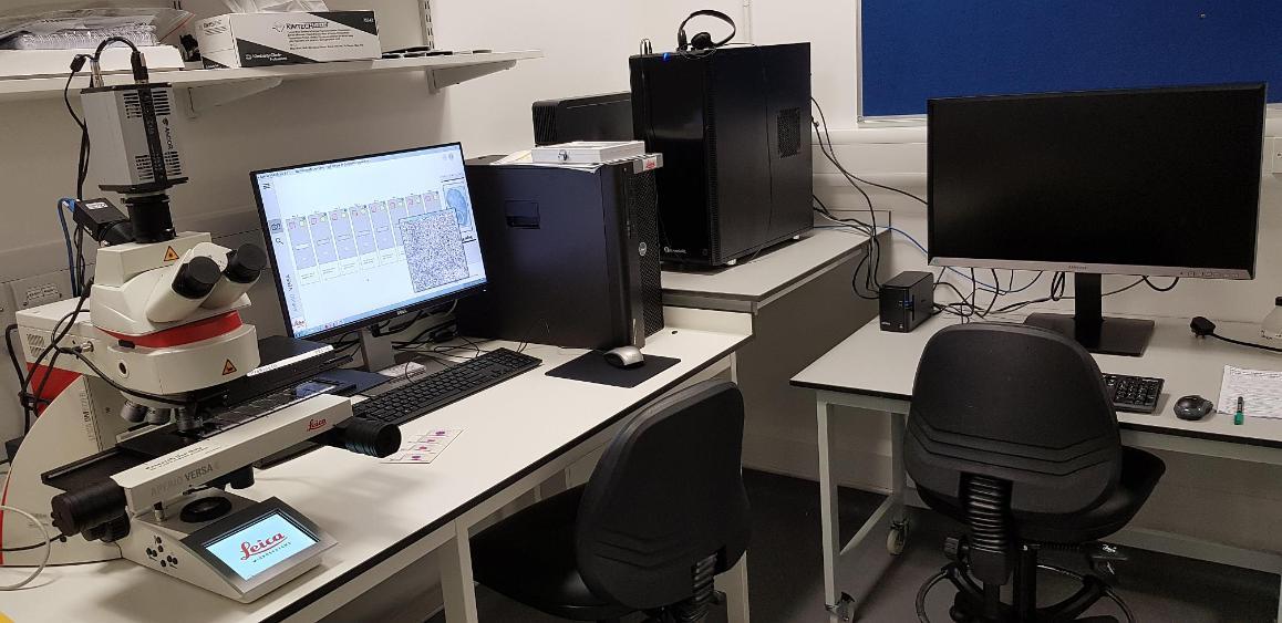
Leica Aperio Versa 8 slidescanner
The Leica Aperio Versa 8 slidescanner allows automated scanning of up to 8 slides, either brightfield or fluorescently stained.
The dichroic filters available are:
|
|
Excitation |
Emission |
Some fluorophores that work |
|
Blue |
350/50 |
460/50 |
Hoechst, DAPI |
|
Green |
482/25 |
525/30 |
FITC, GFP, Alexa 488 |
|
Red (lower end) |
546/22 |
590/33 |
Texas Red, Alexa 555/568 |
|
Red (higher end) |
580/25 |
625/30 |
Alexa 594 |
There are 5 (dry) objective lenses fitted, ranging from 1.25x to 40x.
Image analysis is achieved using Leica Imagescope software.
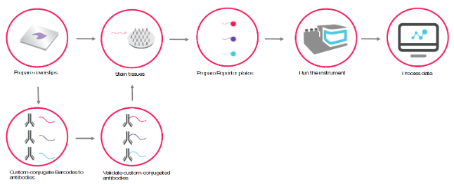
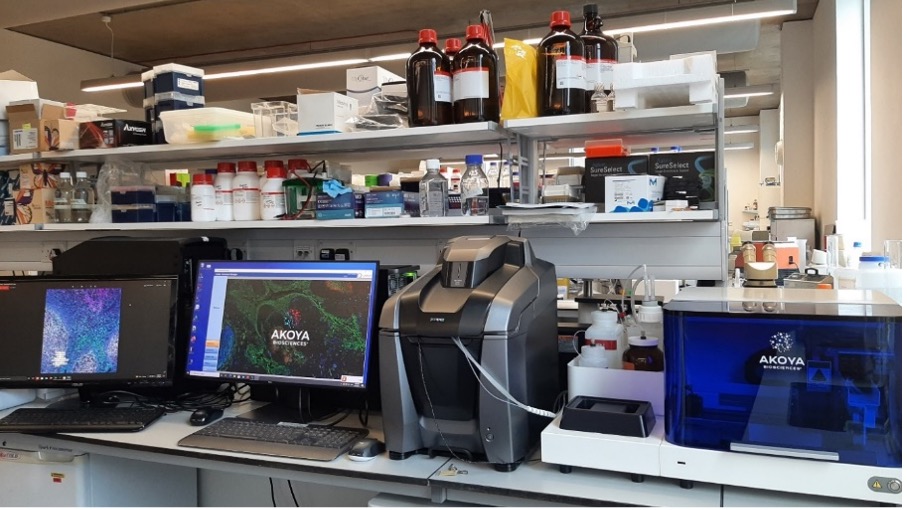
Akoya CODEX Multiplex Imager
The CODEX (Co-Detection by Indexing) technology combines the advantages of single-cell biology and histology. The CVR CODEX is combined with a Keyence BZ-X810 fluorescence microscope enabling highly multiplexed (40+) biomarker analysis in both fresh-frozen (FF) and formalin-fixed paraffin-embedded (FFPE) tissue sections. The combination of high-resolution imaging and high-parameter detection reveals different cell phenotypes, and how they interact and organize across the entire tissue landscape to impact disease pathology and progression.
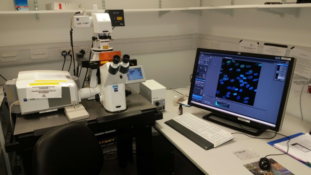
LSM 710 Confocal Microscope
Spectra from different fluorophores can be so similar that they can’t be separated using ‘standard’ PMT detectors used on confocal microscopes. The LSM 710 uses a 32-channel quasar detector to unmix fluorophores even if they have significantly overlapping spectra. ‘Lambda’ mode allows this to be done on previously acquired images (once reference spectra are available) whereas ‘Online Fingerprinting’ facilitates unmixing ‘on the fly’ as images are acquired. Both methods are extremely useful and very quick once set up. The LSM710 has so far been used to unmix 7 fluorophores successfully at the CVR.
The microscope is fitted with the following hardware:
|
Objective lenses |
Laser lines |
Detectors |
|
10x (dry) – DIC ready |
405nm (eg DAPI) |
2 x PMTs |
|
20x (dry) – DIC ready |
458nm (eg CFP) |
1 x spectral detector |
|
40x (oil) – DIC ready |
488nm (eg GFP) |
|
|
63x (oil) – DIC ready |
514nm (eg YFP) |
|
|
100x (oil) – NOT DIC ready |
561nm (eg Alexa 568) |
|
|
|
633nm (eg Alexa 633) |
|