Live-Cell Time-Lapse Microscopy
The CVR has a range of modalities to support time-lapse imaging of unstained or fluorescently tagged/stained live cells:
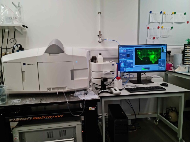
Lightsheet Microscopy
The CVR’s Zeiss lightsheet Z1 microscope is the only Zeiss Z1 lightsheet microscope installed in Scotland. It provides flexibility in the CVR’s capacity for deep imaging. It can gently and quickly image large specimens up to 1cm3, fixed (cleared or aqueous) or live, detailing dynamic biological processes and providing context to higher resolution imaging modalities already located within the CVR such as super-resolution light microscopy and cryo-electron microscopy.
See some of the CVR’s lightsheet-acquired images (link to lightsheet images in showcase)
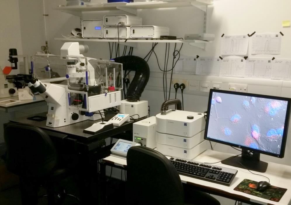
Widefield Deconvolution Microscopy
The Zeiss Axio Observer Z1 is the CVR’s primary system for livecell time-lapse image acquisition. In addition to fluorescence it supports DIC- and phase contrast-imaging of unstained samples. It also has Zeiss’s deconvolution module installed.
Confocal Microscopy
The Zeiss LSM880 is the CVR’s newest confocal microscope. It is enclosed in an environmental chamber so is capable of imaging with both fixed and live samples. Fitted with a highly sensitive GaAsP detector it acquires images with high signal:noise, supporting acquisition of both fluorescent and white light DIC or phase contrast images.
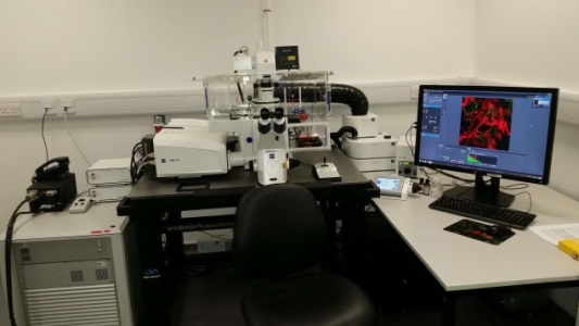
Super-resolution Microscopy
The Zeiss LSM880 as well as working as an excellent confocal microscope is also fitted with an Airyscan FAST module for super-resolution imaging, theoretically providing images with resolution approaching 100nm.
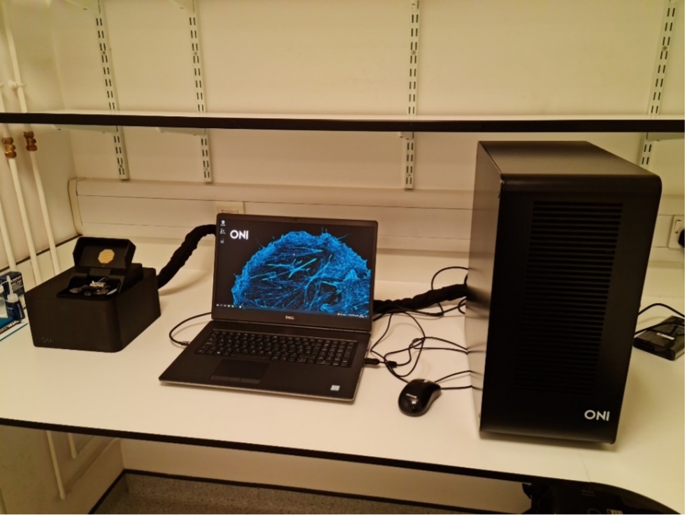
The ONI Nanoimager is the CVR’s newest imager. It has full environmental control to facilitate time-lapse imaging at super-resolution (dSTORM, PALM, single-particle tracking, TIRF, HILO, smFRET).
One of the biggest challenges in pathogen research is their size; in the range of micrometers down to nanometers. Visualizing their propagation and interaction with the host can be extremely challenging. Advances in having better tools to observe and study the architecture, interactions, and drug-targeting effects on such pathogenic agents will bring scientists closer to finding more efficient ways of treating and eradicating devastating infectious diseases. Conventional microscopy techniques, such as confocal microscopy, are limited by the diffraction of light, with individual molecules only resolved at a spatial resolution of 200 nm, at best. In contrast, single-molecule localization microscopy (SMLM) techniques such as dSTORM or PALM allow the visualization of specific biomarkers (proteins, antibodies, nucleic acids, and other biomolecules), their distribution and interactions on biologically relevant scales, achieving resolutions of 20 nm (NB paragraph of text taken directly from ONI website).
Note for local users: The local user pages provide details of equipment curators and locations.