

Short films
This series of short films aims to introduce you to our work and to provide insights into some of our research techniques, partnerships and public engagement work.
Dissection of a Sand Fly Gut : Exploring Leishmaniasis with Dr Hector Diaz Albiter
Leishmaniasis is caused by the Leishmania parasite which is found in many parts of the world including southern Europe, the Middle East, India, North and South America, and Africa. Millions of people are currently at risk, and the distribution is altering as a result of global warming. The parasite is carried by a sand fly vector in a similar way that malaria is transmitted by an infected mosquito.
Some Leishmania species cause unsightly skin ulcers (cutaneous Leishmaniasis) at the site of the sandfly bite that eventually heal on their own, although drug treatment can speed up this process. However, with some cutaneous Leishmaniasis-causing species, there is a risk that several months or years following healing or treatment, the patient will develop an even more disfiguring form of the disease called mucocutaneous Leishmaniasis where the parasite destroys mucous membranes of the face. This involves the parasite travelling from the original site of infection (wherever the sandfly bite occurred) to the nose or mouth. Another form of disease caused by Leishmania parasites is visceral Leishmaniasis, which affects internal organs such as the liver and the spleen and is often fatal unless treated. Here, the Leishmania parasites must travel from the bite site to the internal organs to cause disease.
The parasite has a complex lifecycle. An important part of this lifecycle takes place in the gut of a sand fly. Therefore in order to study this stage in more detail, the parasites must be removed from an infected sand fly. In this short film, Dr Hector Diaz Albiter demonstrates how this procedure is carried out. It is very delicate and highly skilled work – the sand flies are only 2mm in length!
Using Comics to Communicate Research into Parasites
The Centre of Parasitology (previously Wellcome Centre for Integrative Parasitology) has produced a series of comics that explore and explain our research.
Investigating Chagas Disease with Dr Hector Albiter.
Chagas Disease is found in Latin America and is also known as American Trypanosomiasis as it is caused by a parasite from the same family as the parasite that causes African Trypanosomiasis – or sleeping sickness. It is spread mostly by insects known as Triatominae. The insects are found in houses made from materials such as mud, adobe, straw, and palm thatch. During the day, they hide in crevices in the walls and emerge at night when people are sleeping. Because they tend to feed on people’s faces, triatomines have become known as “kissing bugs. ” After they bite and ingest blood, they defecate on the person. The person can become infected if T. cruzi parasites in the bug faeces enter the body through mucous membranes or breaks in the skin. Often, a sleeping person may accidentally scratch or rub the faeces into the bite wound, eyes, or mouth.
There are two phases of Chagas disease: the acute phase and the chronic phase. The acute phase lasts for the first few weeks or months of infection. It usually occurs unnoticed because it is symptom free or exhibits only mild symptoms such as fever, fatigue, body aches, headache, and rash. These symptoms usually fade away on their own within a few weeks or months. However, if the infection is not treated, it can persist and the infected individual can develop more chronic symptoms. During the chronic phase (which may take years to develop) cardiac or intestinal complications can develop which can result in ill health or death. It is estimated that approximately 8 million people are infected with Chagas Disease in Mexico, South and Central America.
Like many parasites, T.cruzi has a complex lifecycle. In this short film, Dr Hector Diaz Albiter, outlines the lifecycle and looks in depth at the vector – the “kissing bug”.
Rotation Project: Toxoplasmosis in Lego by the Harding Lab
The Harding lab’s research will focus on how Toxoplasma acquires and uses iron from its host cells. Iron is an essential nutrient for almost all cell types, from bacteria to the cells that make up our bodies, however it can be difficult to acquire and is toxic in the wrong place. Toxoplasma is the ultimate generalist, it requires a host cell to replicate within, it can grow within any cell type from any warm blooded animal. This means during its lifecycle, the parasite must contend with both high and low iron concentrations and be able to rapidly adapt to its environment.
To decipher the mechanisms involved, we will seek to answer three key questions:
- How does the parasite acquire iron from its host cell?
- How does iron concentration regulate the expression of genes in the parasite?
- Which proteins and pathways are required for the parasite to adapt to changes in the available iron concentration?
Middle Eastern Biology of Parasitism Course (MeBoP)
Comics
Our comics are an important part of our public engagement programme and have made our research accessible to a wide range of audiences around the world. We have developed a series of comics with comic artist Edward Ross and scientist Dr Jamie Hall - a former PhD student.
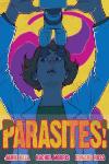
2010 Parasite
Comic artist Rachel Morris designed the cover. The comic proved hugely popular with a range of age groups and continues to be used for public engagement. It gives a brief overview of parasitology, and the parasites we study.
2012 Malaria: The Battle Against a Microscopic Killer
Was again written and drawn by Jamie and Edward with input from artists Tom Humberstone and Luke Pearson. It has been translated into many languages, including Swahili. This comic was supported by Evimalar - a former network of European research groups collaborating on malaria research.
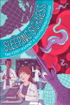
2016 Sleeping Sickness: The Fight Against a Nightmarish Disease
Explores another important parasitic disease that is studied by researchers at the WCIP and highlights the work of the MacLeod Group.
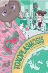
2017 Toxoplasmosis: Unlocking the Secrets of a Mysterious Parasite
Highlights our research on the Toxoplasma parasite and features the work of the Sheiner Group.
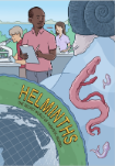
2020 Helminths: The Secret World of Parasitic Worms,
Tells the story of our research on schistosomiasis, a disease caused by a parasitic worm carried in snail vectors. This comic also talks about how these parasites may offer some hope in the understanding and treatment of autoimmune diseases such as rheumatoid arthritis. It features the work of the Lamberton Lab and the Maizels Lab.
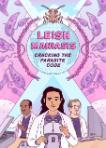
2023 Leishmaniasis: Cracking the Parasite Code
Explores the molecular mechanisms of leishmaniasis and how we can investigate them using CRISPR Cas9 technology. It features the work of the Gluenz Lab, Barrett Lab, Cotton Group and McCulloch Lab.

Comic Translations
Arabic
Catalan
Chichewa
French
German
Hindi
Nepali
Portuguese
Spanish
Swahili
Turkish