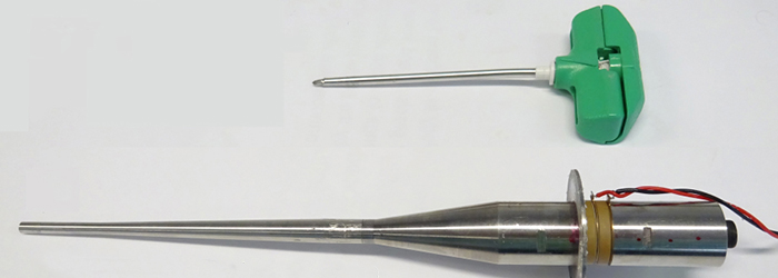
Ultrasonic needle
Bone biopsy is an invasive clinical procedure where a bone sample is taken to aid the diagnosis of a medical condition. Presently, significant force is required to extract the sample which can lead to tissue damage and patient discomfort. A vibrating ultrasonic biopsy needle has been developed to reduce tissue damage and patient discomfort by minimising the force applied.

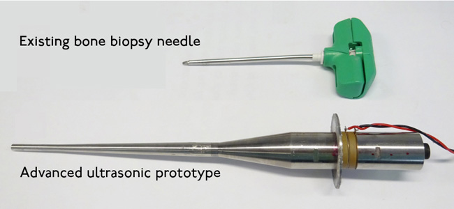
This image shows the the change in the needle design.
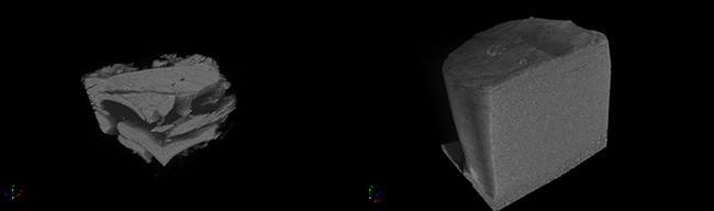
CT scans of hard bone sample show the difference in the two needles. The sample taken with the existing biopsy needle is shortened and twisted (left image). This is consistent with a high force being applied. The sample taken with the ultrasonic needle has a uniform cylindrical profile and shows no evidence of distortion (right image). This is consistent with the application of a small force.
Find out more about Ultrasonics at the University of Glasgow.
Funded by

Partners: