Medical Illustration
Modern Medical Illustration departments only evolved after the inception of the National Health Service. Previously, University technicians and commercial artists had augmented the work done by teaching professionals and clinicians who had produced their own visual aids.
The situation in the 1960s at the Royal Infirmary had evolved into three small units attached to the University Departments of Medicine, Pathology and Surgery and a larger central NHS department situated next to the Pharmacy in the basement of the main building. Two other small departments serving Burns and Plastics at Canniesburn and the Dental School and Hospital completed the Eastern District Group of Medical Illustrators.
A similar pattern of evolution occurred at the Western Infirmary, with a larger central department and smaller, occasionally single handed units serving specialty departments. Illustration units had also been established at the Victoria Infirmary and Southern General Hospital and on the site at Yorkhill of the Maternity and Sick Childrens’ Hospitals. 
The broad concern was to provide a comprehensive service of illustration which served the requirements of the users;
- teaching aids for University lecturers
- clinical records for clinicians
- diagnostic aids in areas such as pathology photomicrography and macrography
- ophthalmic retinal fundus photography and fluorescein angiography.
Pioneers
Robin Callander
Possibly the highest profile practitioner was Robin Callander, co-author with Dr Anne McNaught of Illustrated Physiology, in the 1960’s, the first of a worldwide series of Illustrated medical textbooks.
Enlisting, aged twenty in 1942 in the Royal Air Force, he trained in radio technology and photography and was commended for his work alongside U.S. Army Air Corps personnel in developing and illustrating technical manuals. On leaving the R.A.F. he trained as a physiotherapist and in the early 1950s accepted a post (temporary at first) from Professor RC Garry to provide an illustration service to staff and students at the West Medical Buildings of the University of Glasgow. He quickly established himself as a skilled illustrator and sympathetic collaborator, producing illustrations for many notable text books and publications, including Garven’s Histology.
When the University restructured illustration services he became the inaugural Director of Medical Illustration and continued to influence the practice of Medical Illustration through his work with the Medical Artists Association of Great Britain and the Institute of Medical and Biological Illustration, serving both as Chairman.
Gabriel Donald
Another notable pioneer, based in the Western Infirmary was Gabriel Donald. He had the choice of entering medicine or art; he chose art but ended up working in the life sciences. And medicine itself! After receiving the diploma in art for drawing and painting from the Glasgow Art School, where he was a contemporary of the great Scots-Canadian film animator, Norman McLaren, and Ralston Gudgeon, the celebrated wildlife artist, he undertook further studies, gaining the art teachers' diploma, and taught art at senior secondary school level in the city in two areas of social deprivation.
Called up for service in the Second World War he trained as a pilot officer and was eventually a flight lieutenant with the illustrious Pathfinder Squadron, finally flying Mosquitos deep into enemy territory. It was during the last three months of the war that he was shot down over Germany, making a record-breaking freefall parachute descent. He was ultimately captured, transported by train to a Prisoner of War camp, but subsequently escaped.
His interest in figure drawing at the School of Art and his obvious skill in anatomical illustration led him from teaching to medical art. In this he was following on in the tradition of those early pre-Christian artists who depicted the treatment of arrow wounds on Greek vases through to Leonardo da Vinci's highly detailed life sketches based on a firm understanding of anatomy and physiology.
He was asked to undertake a few drawings by Professor W J R B Riddell of the Tennant Institute of Ophthalmology and made such an impression with Riddell that he left school teaching and was appointed an assistant lecturer in Ophthalmology at the University of Glasgow. This led to his remarkable career in medical illustration and subsequently his appointment as Director of Medical Illustration when the then Western Regional Hospital Board and Glasgow University decided to establish a separate department to cater for both NHS service requirements and the teaching needs of what was to become the largest medical school in the UK.
He illustrated the seminal textbook, The Fundus of the Eye by Ballantyne & Michaelson, and went on to have a long association with Professor Wallace Foulds of the Tennant Institute and with the University's Regius Department of Surgery, working closely for many years with both Sir Charles Illingworth and Sir Andrew Watt Kay.
Gabriel Donald was not only a highly accomplished artist but was an award-winning cinematographer and medical educationist. He collaborated with William Dunn of the Institute of Educational Studies at the University and Sir Edward Wayne of the Gairdner Institute of Medicine in the development of new and experimental methods of clinical teaching, including pioneer work on tape-slide teaching methods, and, in conjunction with Dr Gavin Shaw and the late Bill Brown of Scottish Television, broadcast continuing medical education programmes in Scotland.
He was chairman of the Medical Artists' Association of Great Britain for more than 20 years and was the founding co-chairman, with Dr Peter Hansell, of Westminster Medical School and the University of London's Institute of Ophthalmology, of the Institute of Medical and Biological Illustration in 1968. He was appointed MBE in the New Year's Honours List in 1980. He was an adviser to the Royal College of Physicians and Surgeons of Glasgow and, outside of work, his interest lay in landscape painting and gardening and was a former Visitor of the Incorporation of Maltmen of the Trades House of Glasgow.
Royal Infirmary
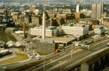
During the 1970’s a steering committee under the stewardship of Dr J Killoch Anderson, (the hospital superintendent) outlined plans for a new Department of Medical Illustration, incorporating all of the existing staff, to be housed in the ground floor of the new University Tower (Queen Elizabeth Building) which was planned for the recently cleared site on Alexandra Parade. Some preliminary work had been done by Mr Joe Devlin, (subsequently head of the service at Yorkhill and Sick Childrens’ Hospital) and Mr Alan McIlroy (head of the service at the Southern General Hospital), identifying the style of layout, service provision and basic equipment requirements.

Contact through the Institute of Medical Illustrators in Scotland (IMIS), and after its incorporation into the UK wide Institute of Medical and Biological Illustration (IMBI), with other graphic and photographic professionals helped shape the layout and service provision aspirations of the new unit.
A wish list of essential equipment was drawn up and University funding (usually approaching the first of April spending deadline) was released in tranches and invested in capital items providing the core of the service.
Under a new head of service the department was able to provide a comprehensive range of regular clinics in
- Ophthalmology (producing diagnostic retinal fundus images and Fluorescein Angiography sequences)
- Dermatology (including visual recording of clinical trial results and diagnostic clinical comparison images)
- Oral medicine
- General medicine (including specialist Endocrine clinics)
- and a sophisticated Surgical response service to record both elective and unusual or unexpected events across the full surgical spectrum.
The Accident and Emergency department at “Gate” provided a vast range of trauma which was recorded in measurable and repeatable formats.
The department adopted the “Westminster” method of standardisation; all clinical images were recorded at a series of fixed scales against a standard background colour. Apple green in the studio (a colour which rendered skin tone in its most natural appearance) and theatre drape green in clinical and surgical locations. This allowed for accurate clinical measurements and direct comparison as application of these techniques mirrored those in many departments throughout the UK.
Advances in audio-visual technology were quickly adopted and as open heart surgery and vascular surgery advanced, video technology allowed these techniques to be shared as teaching aids.
Alongside the huge range of clinical and surgical work undertaken the laboratory techniques behind many of the specialties could now be adequately visualised for teaching and training. Video programmes allowed detailed imaging of practises pertaining to every discipline for a fraction of the cost of cine production.

The graphic artists continued to provide illustrations as teaching aids, illustrations for learned journals and text books and graphic content for “Poster Presentations”. The changing nature of technologies were quickly reflected in the design and content of teaching presentations. Simple type from manual typewriters or dry transfer lettering on graphs and charts were initially reproduced as negative images, white text on a black background.
By the late sixties coloured diazochrome slides, white against a blue or purple background and hand tinting of slides was popular, allowing complex data to be presented in a clear comprehensive format. An inexpensive and portable system of poster production was evolved, comprising the use of lightweight ticket card with the text and images attached, capable of being rolled and carried in a cylindrical tube. These were provided with velcro tabs and could be simply unrolled and attached to the exhibition stands provided. This was to replace the earlier style of heavy rigid board which was expensive and relatively awkward to transport, particularly by air.
With the introduction of computer technology, many of the previous limitations on the production of good visual aids disappeared. Sophisticated full colour illustrations could now be produced straight from the screen and clinical images could be imported and manipulated or annotated as required. This also signalled the eventual contraction of the profession, as many younger computer literate medical professionals preferred to produce their own material, a trend which continues with the adoption of smart phones, capable of producing high definition images and video and sharing these instantly.
Fraser Speirs

