

In situ structure of virus capsids within cell nuclei by correlative light and cryo-electron tomography
Published: 20 November 2020
Researchers at the Institute's Centre for Virus Research have used cutting-edge imaging techniques to reveal, for the first time, the structure of the herpes virus within the cell.
Researchers at the Institute's Centre for Virus Research have used cutting-edge imaging techniques to reveal, for the first time, the structure of the herpes virus within the cell.
The Herpes family of viruses are responsible for a range of diseases such as cold sores, chickenpox and in some cases cancer, and cause infection by releasing their DNA into cells.
This DNA is tightly packed within a protective protein shell called a 'capsid', and structural studies often involve isolating virus or cellular proteins from the cell and imaging them in purified form.
However, visualising virus and protein structures within the cell is crucial to understand detailed mechanisms of infection and disease.
The researchers used Nobel-prize winning technology Cryo-electron microscopy, Cryo-electron tomography and advanced computational methods to reveal the structure of the herpesvirus capsids inside cells.
This work revealed the shape of the highly symmetrical capsids and provided insights into their interaction with associated viral tegument proteins that are critical for infection.
Dr Swetha Vijayakrishnan, lead author of the study, recently published in Nature Scientific Reports, said: "It is common to image virus outside of living cells, in an 'in vitro' state.
"Determining structure within cells, i.e.'in situ', is extremely challenging. We used state-of-the-art light and Cryo-electron tomography imaging coupled with high-end computer processing to reveal the structure of herpes virus capsids within the cell.
"Cells are the building blocks of all living organisms and their interior is extremely crowded and complex.
"This study allows us to peer into the cell, to visualise how viruses and proteins look and work as well as potentially how they interact with one another.
"We hope this work will eventually enable looking at how virus or proteins are affected by therapeutic treatments inside the cell and how this correlates with disease states."
In situ structure of virus capsids within cell nuclei by correlative light and cryo-electron tomography
- Swetha Vijayakrishnan, Marion McElwee, Colin Loney, Frazer Rixon & David Bhella
- Scientific Reports, volume 10, Article number: 17596 (2020)
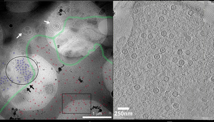
Image (top): (L) EM imaging of refrozen cell sections using, for easy visualisation, segmentation of the image using Photoshop is shown annotating probable edges of the nuclear membrane (green), nuclear capsids (red) and cytoplasmic capsids (blue); (R) Electron tomography of nuclear and cytoplasmic HSV capsids.
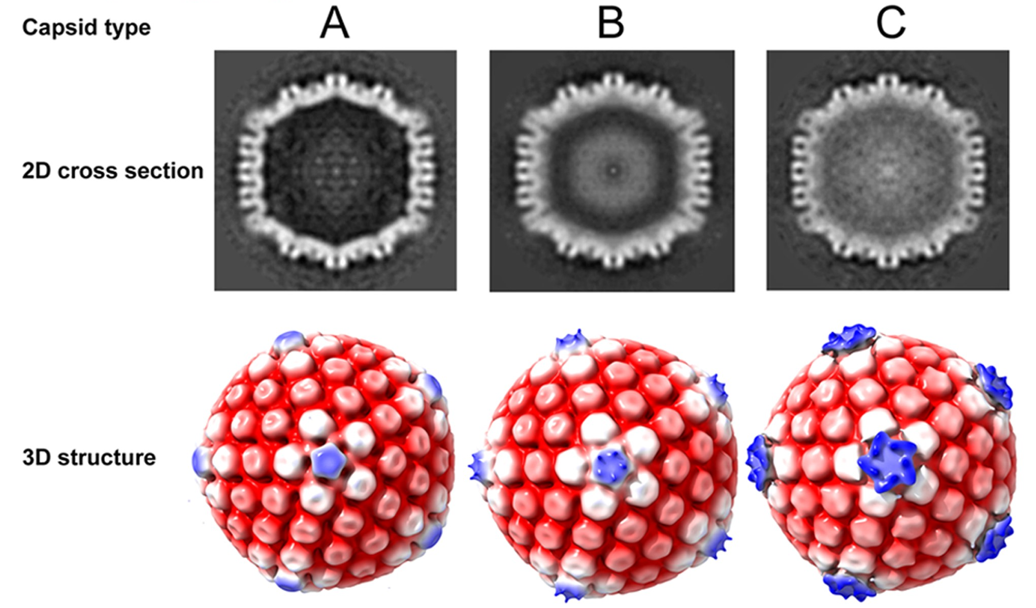
Image (centre): Determination of the 3D HSV capsid structures from within the nucleus. 3D structures of the three types of nuclear capsids, A-, B- and C-type were determined by subtomogram averaging. 2D cross-section images clearly indicate density differences in the interior of the capsid between the three types. Density relating to scaffolding proteins and DNA is seen inside the capsids within the B-type and C-type, respectively, while none is present inside the empty capsid shell of the A-type. The 3D structures obtained have been radially coloured according to distance (row 2) at low thresholds of density and are shown along the fivefold axis of icosahedral symmetry. Pronounced pentaskelion density is observed above the pentons only in the C-type capsids.
First published: 20 November 2020