

3I’s Imaging Competition
Published: 25 May 2018
The winner of this year’s Imaging Competition was announced on Monday 21 May. Well done to Ashley Roberts from the Centre for Virus Research (CVR), who was awarded the first prize of a £250 travel grant.
The winner of this year’s Imaging Competition was announced on Monday 21 May. Well done to Ashley Roberts from the Centre for Virus Research (CVR), who was awarded the first prize of a £250 travel grant.
A special mention also goes to Colin Crawford and Floriane Almire who secured second and third place, respectively.
The quality of this year’s entries was really impressive, so thank you and well done to everyone who took part.
Ashley Roberts (1st):
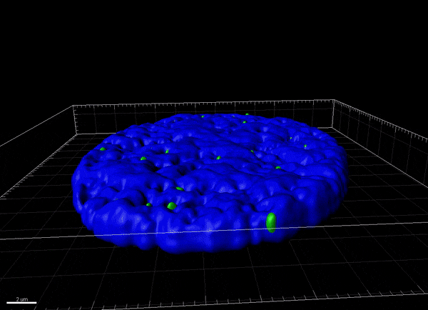
Ashley's entry shows a 3D reconstruction of a cell nucleus (blue) infected with Herpes simplex type 1 which causes cold sores in humans. Viral genomic DNA (red) can be seen encased within a PML-nuclear body which contains cellular restriction factors including PML (green) that act to prevent the initiation of infection.
Technical Summary
A Zeiss LSM 880 confocal microscope was used to capture z series images with the 63x Plan-Apochromat oil immersion lens (numerical aperture 1.4), the 405 nm, 488 nm, 543 nm, laser lines and the PMT and GaAsP detectors under Airy scan conditions. Eighty images were captured at 0.2um intervals using the Airy scan detector and then deconvolved using the Airy scan processing in Zen Black (Zeiss). Deconvolved images were then further processed using Imaris (Bitplane) software to render three dimensional image reconstructions.
-----------------------------------------------------------------------------------------
Colin Crawford (2nd):
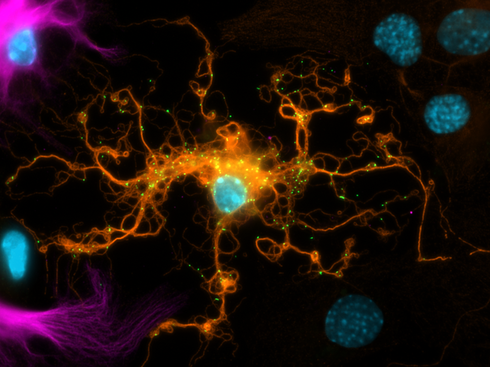
Colin's entry shows a lab-grown oligodendrocyte in the centre of the image. In the brain, these cells provide insulation and support to neurons. We’ve visualised the cell's skeleton (orange) and can see peroxisomes (green), small organelles, which move along the cell's skeleton "tracks". We also see astrocytes nearby (violet).
Technical Summary
This image was captured on a Zeiss Imager.M2 fluorescence microscope using a 63x oil objective. Visualised here is DAPI (blue), mEOS2-labelled peroxisomes (endogenous labelling with peroxisome targeted mEOS2; green), β-tubulin IV (Alexa Fluor 568; orange), and glial fibrillary acidic protein (GFAP; Alexa Fluor 647; violet).
-----------------------------------------------------------------------------------------
Floriane Almire (3rd):
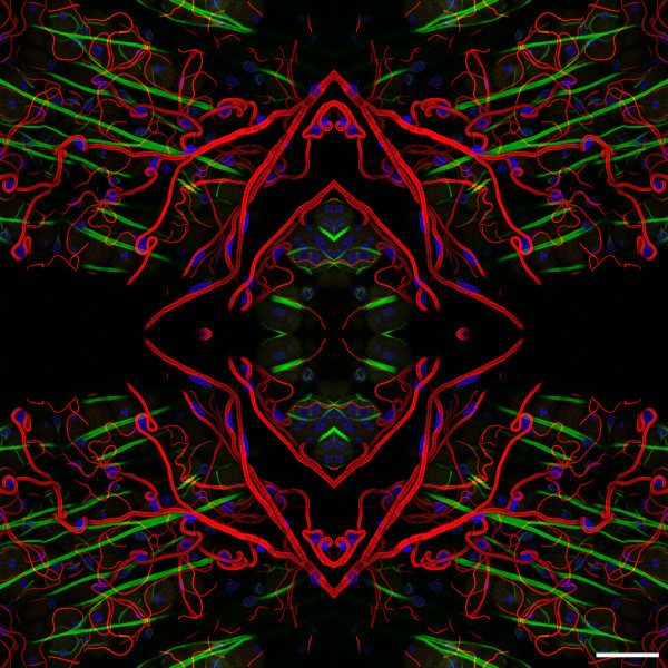
Floriane's entry is composed of four identical images, which are symmetrically organised. It shows the tracheal network (or respiratory system) of the gut in Aedes aegypti female mosquito, vector of arboviruses such as ZIKV. The gut is immunostained for tracheae (red). Nuclei are stained with DAPI (blue) and the actin is stained with Phalloidin488 (green).
Technical Summary
The image was acquired on a Zeiss LSM 710 inverted confocal microscope with a 40x oil-immersion objective. Scale bar: 40μm.
First published: 25 May 2018