

Excellence in imaging
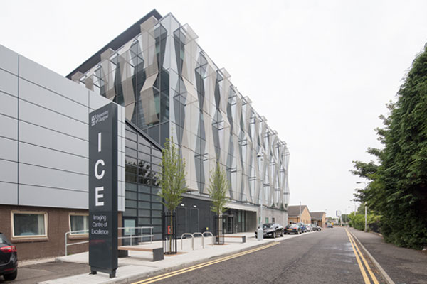
The University of Glasgow’s new Imaging Centre of Excellence (ICE) is a pioneering hub developing ground-breaking scientific research into real benefits for patients with chronic disease.
The state-of-the-art £32million ICE building offers greatly enhanced medical imaging technology, most notably in the area of brain imaging, and is connected to the £1billion Queen Elizabeth University Hospital (QEUH) in Glasgow, the largest acute hospital in Western Europe.
ICE is supported by the UK Government and the Medical Research Council, as part of the Glasgow City Region City Deal, the UK Research Partnership Investment Fund, the European Regional Development Fund and the Dr Mortimer and Theresa Sackler Foundation.
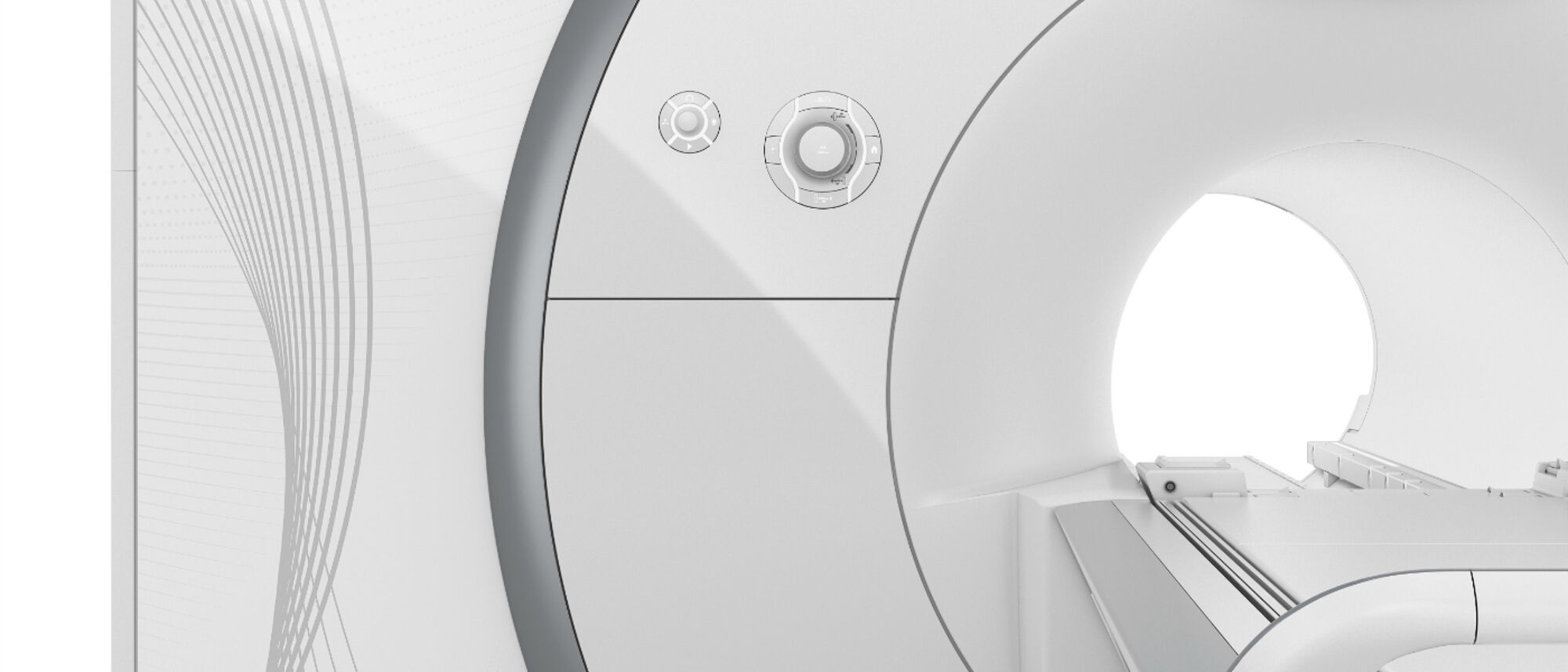
The Centre’s facilities include an ultra-high resolution 7-Tesla (7T) MRI scanner, the only one of its kind in Scotland and the first in the UK to be fully integrated within a clinical site.
There are only a handful of these machines worldwide, placing Glasgow at the forefront of medical imaging capabilities, as a driving force for precision medicine.
Improving understanding of chronic diseases
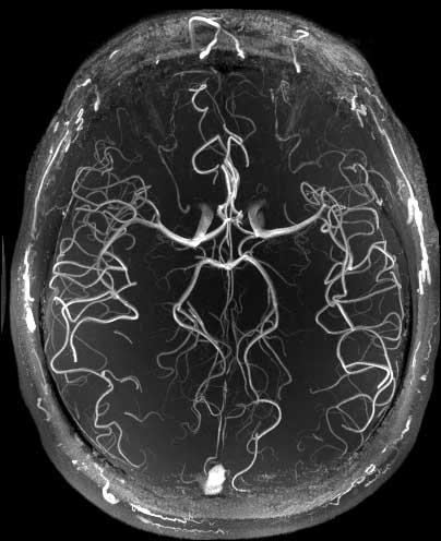
The 7T scanner allows scientists and clinicians to study the human body in greater resolution than ever before, hugely benefiting patients across the UK and beyond.
“The superconducting magnets produce a magnetic field more than twice as strong as those in current clinical scanners”, says Professor Keith Muir, SINAPSE Chair of Clinical Imaging (Stroke and Brain Imaging).
“The high magnetic field of the 7T scanner allows extremely high resolution scanning of the body, potentially letting us see structures down to 1 millimetre in size or even smaller.
“This technology opens up the possibilities for new clinical and basic neuroscience research, and has great potential for aiding our understanding of diseases such as stroke, vascular dementia, brain tumours, Parkinson’s, Alzheimer’s, epilepsy and multiple sclerosis.
“For my specialism in stroke, the scanner will allow us to produce detailed images of the damage caused by stroke and give us the capability of visualising the very small vessels responsible for many strokes,” Professor Muir explains.
“These devices are already being used for research into the brain. But so far we’ve only scratched the surface of what these pieces of equipment can do.”
The medical research being carried out on the 7T scanner could lead to new ways of diagnosing diseases, as the machine will not only speed-up scientific research but will also directly help patients.
The forefront of stroke diagnosis and treatment
Professor Muir is using the scanner in clinical research trials to devise advanced imaging methodologies to improve prognosis in early stroke.
His research seeks to apply advanced imaging methods – including the opportunities opened up by 7T MRI - to the treatment of acute stroke. Clinical studies can exploit the range of imaging equipment and analysis facilities associated with the ICE building.
“What sets Glasgow’s 7T scanner apart from those at other sites in the UK – and indeed internationally – is that it is integrated into a major acute hospital. Most other facilities can only handle patients who can walk into a university research building, but we can bring patients in their beds from the hospital directly into the ICE building to use the scanner”. Professor Keith Muir
Stroke affects over 125,000 people each year in the UK and leaves at least 50% disabled. Treatment of stroke caused by a blockage in a blood vessel (ischaemic stroke) with clot-busting drugs improves the chances of good recovery, but must be given within 4.5 hours of onset.
Currently only a small proportion of patients who arrive in hospital within 4.5 hours are treated. This is largely due to uncertainty about diagnosis and concerns about risk of bleeding associated with clot-busting medication.
Patients with mild or improving symptoms in particular are often not treated because of uncertainty about relative risks and benefits. However, around one third of these patients go on to be significantly disabled.
Using imaging to improve treatment
Professor Muir aims to use imaging as the mechanism of stratification - rather than ‘time since onset of stroke’, or severity of symptoms - and is the Principle Investigator of the Penumbra and Recanalisation Acute Computed Tomography in Ischaemic Stroke Evaluation (PRACTISE) trial.
The PRACTISE trial is investigating the value of additional computed tomography (CT) scanning that gives information on the blood vessels (CT angiography) and blood flow to the brain (CT perfusion) by undertaking a randomised trial.
Extra scans are done in the same scanner and involve some extra radiation, injections of a contrast dye, and some extra time to acquire, process, and interpret.
This may expand treatment options for patients not typically eligible for clot-busting treatment, e.g. those with mild stroke (who often deteriorate later without treatment, but are often not treated due to concern about side effects), while reducing any risk for those who are unlikely to benefit e.g. those who have no salvageable brain tissue and might be more at risk of harm from treatment.
The extra scans may allow better treatment decisions for patients by increasing diagnostic certainty and by better assessment of stroke severity.
Professor Muir’s team have also led the EU-funded WAKE-UP trial for the UK. The trial aims to provide a new safe and effective treatment option for the 25% of acute stroke patients who wake with stroke symptoms already present, or whose stroke came on without being witnessed.
The WAKE-UP consortium brings together leading stroke researchers from European academic institutions, highly specialised enterprise partners, and patient organisations providing a wide range of clinical and scientific expertise in stroke, image processing, and the conduct of clinical trials.
The trial is using MRI to identify individual patients who may fall into the time window for clot-busting drug treatment when the timing of symptom onset is unknown.
A pioneering theronastic tool
Professor Muir’s research is also developing an improved diagnostic tool for acute stroke that has therapeutic potential, known as theranostic.
The research, led by Aurum Biosciences - a spin-out from NHS Greater Glasgow & Clyde and the University of Glasgow - has investment of over £3million to develop the theranostic stroke imaging tool.
The technique is a contrast MRI imaging method referred to as Glasgow Oxygen Level Dependent (GOLD) technology.
GOLD uses oxygen together with an injectable perfluorocarbon-based oxygen carrier as an agent to enhance the signal from oxygen uptake by the brain.
The technique allows mapping of the areas of the brain where there might be salvageable ischaemic tissue (penumbra) after a stroke by seeing whether tissue can take up oxygen or not. The extra oxygen carried by the perfluorocarbon molecules greatly strengthens the signal.
Delivering extra oxygen has the added benefit that it may sustain the ischaemic brain tissue. Current treatment for stroke such as clot-busting therapy is effective only within a very short time frame of a few hours, so any ability to extend this may be beneficial. It may also be safe enough to administer to stroke patients and suspected stroke patients pre-emptively in the ambulance, to help delay deterioration.
Continuing to give oxygen and oxygen carrier after the scan could also represent a new therapy for stroke, and University of Glasgow animal-based research has provided positive data to confirm this.
Clinical studies are currently getting underway, and in partnership with Aurum, University of Glasgow researchers are working to develop these significant life-saving benefits for patients and healthcare providers.
Collaboration and innovation at ICE
The ICE building, housed within the Clinical Innovation Zone, has been designed to encourage industry, University and NHS collaboration by bringing imaging specialists from a broad range of disciplines together to work on the same site.
“Previously we were all working in separate places and were often faced with the same challenges when it comes to working out how to analyse images. A problem that might take me many months to figure out could be perfectly routine for someone else working in a different discipline – but I might not see that person until I bump into them at the next international conference.
Bringing together researchers into the same building helps us to share our knowledge with each other. Hospitals are already used to radiologists crossing multiple specialist fields, so we’re extending that idea into medical research too.” Professor Keith Muir
This approach aims to transform the management of chronic diseases globally by accelerating biomedical research through open innovation approaches, resulting in high quality health care provision.
Professor Muir and his team will be working with the companies that design and make the hardware and software for the 7T scanners so that they can help to expand the machines’ range of services from the clinical research sphere into the diagnostic arena.
The work taking place in the ICE building is destined to change clinical practice and improve the treatment of chronic diseases like acute stroke, helping patients across the UK and beyond.