

Smart Stent: Minimally invasive therapy for cardiovascular problems
Published: 8 August 2025
Mahmut Talha Kirimi, Steve L. Neale and co-workers show that selectively metallising a non-conductive stent can create electrodes able to detecta build-up of material within and around the stent
Many cardiovascular problems stem from blockages that form within the vasculature and often treatment includes fitting a stent through percutaneous coronary intervention. This offers a minimally invasive therapy but re-occlusion through restenosis or thrombosis formation often occurs post-deployment. Research is ongoing into the creation of smart stents that can detect the occurrence of further problems. For this purpose, a smart stent was created through selective sputtering and gold electroplating (Figure 1).

Initially, the stents were cleaned in an ultrasonic bath at the James Watt Nanofabrication Centre using acetone, IPA, and methanol for 5 minutes, followed by rinsing with reverse osmosis water and drying with nitrogen. During sputter coating, an Agar Sputter Coater was used in cycle mode, where the complete sequence of flush, coat, and vent was automatically controlled by the system. The sputter coater applied a coating of 54 nm gold:palladium (80:20 ratio) that established the seeding layer required for electroplating (Figure 2). During sputter coating, stent was placed on a mask that blocked sputtering to certain areas around the stent through shadow masking, a technique that can be applied to any stent. This stent was then gold electroplated to improve its overall conductivity to be able to be used as a sensor.
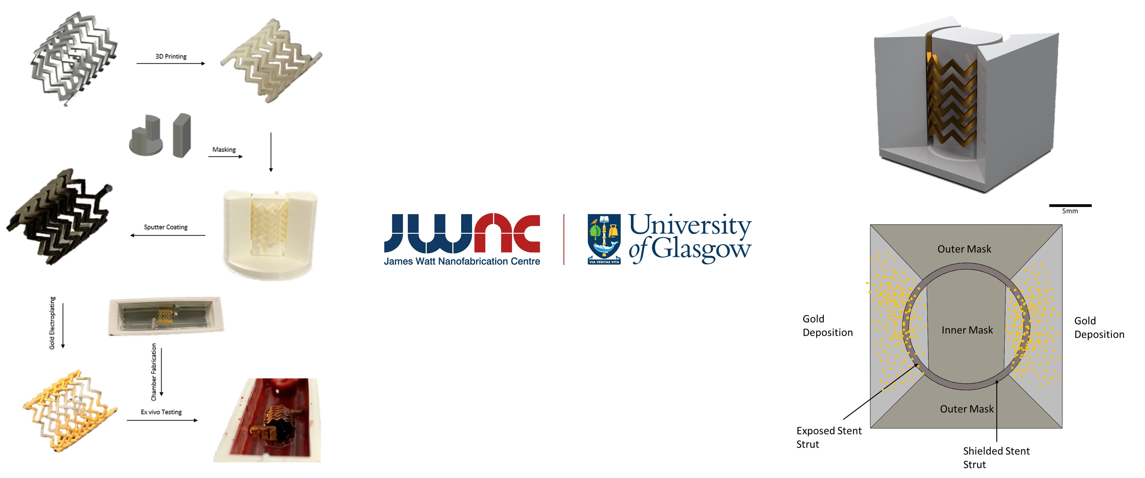
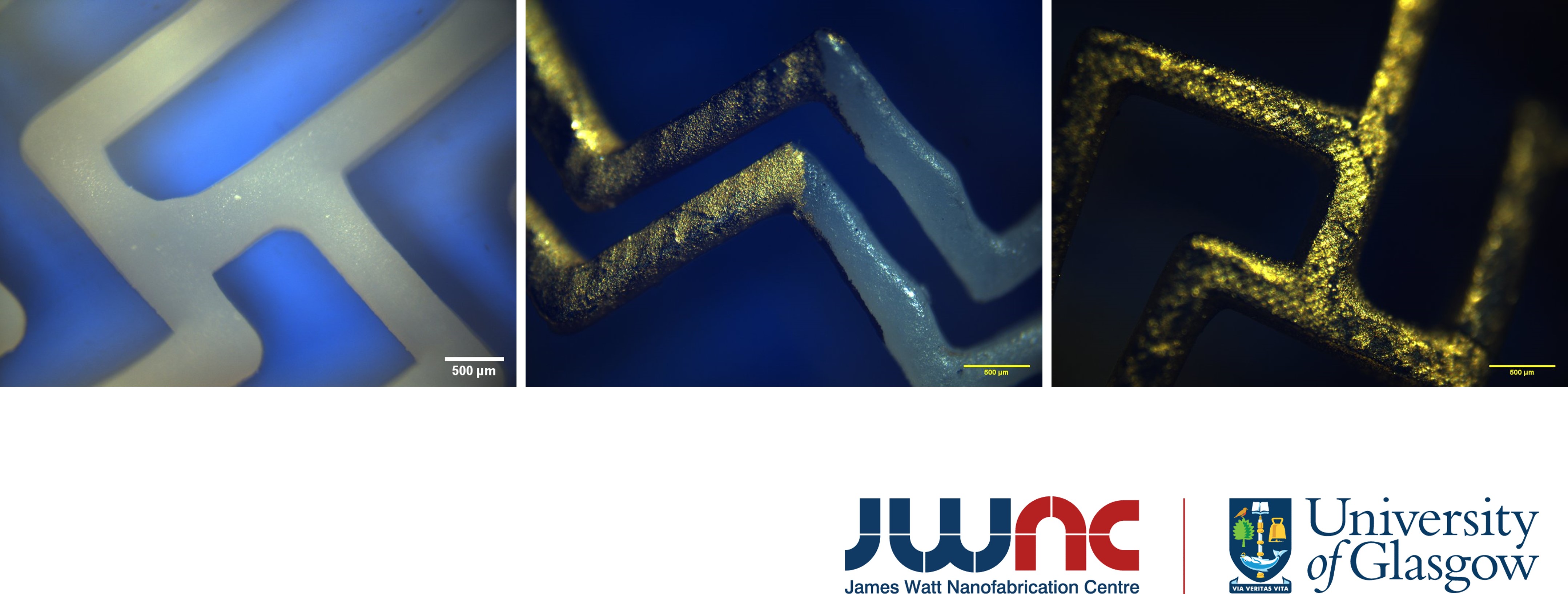
A gold, non-cyanide electroplating solution (ECF 60 ready-to-use solution, Gold Solutions Plating, UK) was warmed to 50°C and agitated using an electronic mixer for approximately one hour in a beaker within a water bath. Once the solution reached the desired temperature, the stent was connected to a power source by threading wires through its struts, submerged in the electroplating solution, and plated for 10 minutes under 2 V and 20 mA settings per conductive half (Figure 2). After electroplating, a multimeter was used to ensure continuity across the entire stent and the separation between conductive and non-conductive parts of the stents were observed under light microscope (Figure 3).
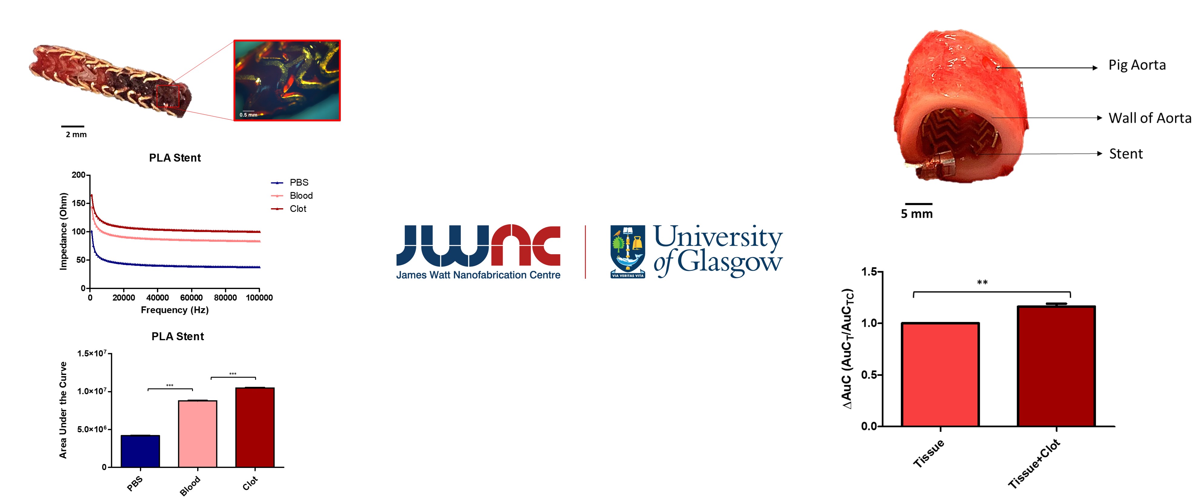
The whole stent sensor was then tested in different conditions and showed that the stent can reproducibly sense the presence of clot showing a 16% ±3% increase in impedance which is sufficient to reliably detect the clot when surrounded by explanted aorta (one sample t-test, p = 0.009, n = 9). It is demonstrated that this approach can be extended beyond the 3D printed prototypes by showing that it can be applied to a commercially available stent, and it is believed that it can be further utilized by other types of medical implants which has the potential to improve quality of life of many patients suffering from cardiovascular problems (Figure 4).
First published: 8 August 2025