

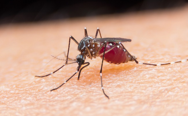
Accumulation of infected red blood cells key to development of cerebral malaria
Published: 19 April 2021
White adipose tissue, or white fat, plays a fundamental role in the development of cerebral malaria in mouse models and humans
White adipose tissue (WAT), or white fat, plays a fundamental role in the development of cerebral malaria in mouse models and humans, according to a new study led by Harvard T.H. Chan School of Public Health scientists in collaboration with the University of Glasgow and an international team of researchers.
The study details the process by which red blood cells infected with malaria-causing Plasmodium parasites are sequestered in small blood vessels throughout WAT, where they stimulate the production of the hormone leptin via a well-studied nutrient-sensing pathway known as mTORC1.
The study was published in Science Advances on March 24, 2021. Among the scientists who contributed to the study was Harvard Chan School’s James Mitchell, who passed away in November 2020, before the study was published.
The findings build on previous research from the group in 2015 that showed leptin production is critical in driving cerebral malaria development and suggested the possibility that mTORC1 inhibitors such as the FDA-approved drug rapamycin could be a viable treatment option. The new findings underscore the potential of rapamycin and similar drugs, and the study may also aid in identifying other treatments as well as in the development of tools to diagnose cerebral malaria and determine the severity of infection.
“It is intriguing that in addition to the brain, infected red blood cells accumulate preferentially in subcutaneous adipose tissue of patients with cerebral malaria. Our study implicates this mechanism in the development of severe disease and opens new therapeutic avenues,” said co-corresponding author Pedro Mejia, who contributed to the research as a Yerby Postdoctoral Research Fellow in Mitchell’s lab at Harvard Chan School. “Professor Mitchell envisioned detecting parasites in this tissue with minimally invasive tools to help discriminate between cerebral and non-cerebral malaria patients and guide medical care in endemic areas. We will keep working on his vision.”
While the group had previously identified that the production of leptin, which is secreted by WAT, drove the development of cerebral malaria in mice, the process was not well understood. To determine whether the sequestration of infected red blood cells into the adipose tissue was essential for disease development, the research team ran studies on several mouse models using several strains of P. berghei parasites that had different capacities for sequestering in adipose tissue.
When they used parasite strains known to sequester in adipose tissue, the researchers found higher leptin levels and a higher lethality in mice. When they used parasite strains that did not sequester in adipose tissue, leptin production was lower and the mice did not develop cerebral malaria. The researchers determined that leptin production in white fat cells was regulated by mTORC1.
To further understand the possible implications of the findings for human health, the researchers analyzed tissue samples from pediatric patients who died from cerebral malaria. They found that the sequestration of infected red blood cells specifically in subcutaneous fat, tissue leptin production, and mTORC1 activity were present and positively correlated with cerebral malaria. The researchers did note, however, that the human serum tissue samples did not indicate that P. falciparum increased systemic leptin levels as observed in mice.
“Exploring the link between adipose sequestration and leptin production proved challenging, but Professor Mitchell was relentless and helped push us to test as many parasitic models and mice models as needed in order to conclusively prove our model. The mechanistic insights on the role of the adipose tissue in the development of cerebral malaria not only consolidate our proposed treatment strategies with mTORC1 inhibitors, but also provide novel insights into how to detect patients likely to develop this often lethal form of malaria,” said co-corresponding author José Humberto Treviño-Villarreal, who conducted the research as a research associate at Harvard Chan School and is currently a professor of endocrinology and molecular metabolism at Mexico’s Universidad Autónoma de Nuevo León.
Co-author Matthias Marti of the University of Glasgow lauded the joint effort required to complete the research. “This is an exemplary collaborative effort using both rodent malaria models and human autopsies, and it was led by my dear friend and outstanding scientist, Professor Mitchell, who is sorely missed,” he said.
Other Harvard Chan School researchers who contributed to this study include Mariana De Niz, Elamaran Meibalan, Alban Longchamp, Justin Reynolds, and Danny Milner. Other institutions affiliated with this research include the University of Glasgow, the University of Bern, Brigham and Women’s Hospital, Harvard Medical School, University of Lausanne, Indiana University School of Medicine, Makerere University, the Pasteur Institute, the Bernhard Nocht Institute for Tropical Medicine, and Michigan State University.
Funding for parts of this study came from NIH grant R01 NS055349, NIH grant R01AI034969, and the European Research Council.
“Adipose tissue parasite sequestration drives leptin production in mice and correlates with human cerebral malaria,” Pedro Mejia, J. Humberto Treviño-Villarreal, Mariana De Niz, Elamaran Meibalan, Alban Longchamp, Justin S. Reynolds, Lindsey B. Turnbull, Robert O. Opoka, Christian Roussilhon, Tobias Spielmann, C. Keith Ozaki, Volker T. Heussler, Karl B. Seydel, Terrie E. Taylor, Chandy C. John, Danny A. Milner, Matthias Marti, James R. Mitchell, Science Advances.
Enquiries: ali.howard@glasgow.ac.uk or elizabeth.mcmeekin@glasgow.ac.uk / 0141 330 6557 or 0141 330 4831
First published: 19 April 2021
<< April