

FLUORESCENCE AND BIOLUMINESCENCE PLATE READERS
BMG Labtech Instruments.
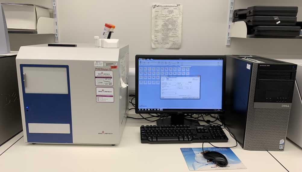
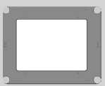
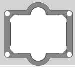
PHERAstar FS
Our PHERAstar FS instrument is a fast reading multifluorescence plate reader and offers the highest spectral emission detector sensitivity. Dye excitation is achieved using a high intensity Xenon Flash Lamp or a solid state N2 UV laser, (e.g. time resolved FRET or BRET assays). Matched green or red sensitive photomultiplier tube detectors are available for optimal detection of green and red dyes. This reader is also equipped with an ultra-fast UV/Visible spectrometer for quantifying absorbance, (220-1000 nm, maximal spectral resolution possible is 1 nm). The system also has on-board reagent injectors for performing real time fast kinetic assays and temperature control regulation is available for performing live cell measurement at 37 degrees C°. Sequential or simultaneous dual emission measurements can also be quantified. Pherastar can read from 6 to 1536 well plates and multiplate readings from different plates can be measured by inserting the plates into a stacker. Another useful feature is cell layer well scanning for multiple read measurements within a well. Well scanning can be performed on 6 to 1536 well plates. Plates can also be automaticall shaken priopr to reading and the instrument has the capability to read from the top or bottom of the microplate well. Automatic focal height z adjustment (0.1 mm resolution) is also available to optimise collection of emission light from the multiplate well. The instrument is located within the ARC building on level 4, room 449. To use the instrument contact Dr John Pediani using the email address below:
A photograph of the PHERAstar FS instrument is shown below:
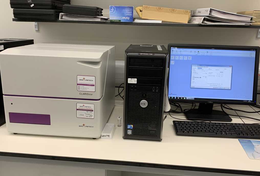
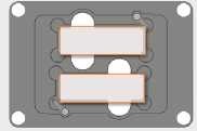
Clariostar
The Clariostar multimode microplate reader is equipped with two tuneable LVF monochromators. Excitation lamp source is a high intensity xenon flash lamp. The LVF monochromator downstream from the flash lamp is tuneable for custom generation of excitation light to excite your dye optimally. The other one is used for custom spectral collection of emission light. A linear variable dichroic mirror is used to efficiently separate excitation light from emission light. LVF monochomator has a linear variable long pass slide and short pass slide which form the rising edge and falling edge of a bandpass filter. Moving the long pass slide and short pass slide to their optimal position creates a filter with a distinct variable peak wavelength and bandwidth. This novel design ensures high transmission of emitted signals with excellent blocking of stray light. Both LVF monochromators have tuneable wavelength selection, (range = 310 to 850 nm) and bandwidth control, (range from 8 to 100 nm), for optiomal excitation and detection of multifluorescence signals emitted from UV-, Visible-, or far Red fluorocrhome dyes. The tuneable design allows weak dyes which have a poor quantam yield to be more efficiently excited by increasing the excitation bandwidth and vice versa bleedthrough of unwanted signals can be reduced by decreasing the bandwidth. The system also has an ultra-fast spectrometer, (<1 sec/well), for performing UV/Vis absorbance measurement (220 to 1000 nm). On-board reagent injectors for performing real time fast kinetic assays and temperature control regulation is available for performing live cell measurement at 37 degrees C°. Automatic focal height z adjustment (0.1 mm resolution) is also available to optimise collection of emission light from the multiplate well.
Please note this instrument has not been fitted with a UV/Visible spectrometer so absorbance measurement is not possible.
The instrument is located within the ARC building on level 4, room 449. To use the instrument contact Dr John Pediani using the email address below:
A picture of the Clariostar instrument is illustrated below:
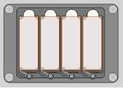
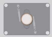
Omega Polarstar
Our Omega Polarstar microplate reader is equipped with high energy Xenon Flash Lamp and has an 8 position excitation and emission filter wheel for sequential multi excitation and emission. System is equipped with two side window photomultiplier tubes, (spectral range 240-900 nm), for sequential or simultaneous dual emission recording of emitted signals (e.g. fluorescence intensity, FRET, fluorescence polarisation, Luminescence (flash and glow) - including BRET and Alphascreen). Bottom read absorbance measurements over the wavelength range 220-850 nm with selectable spectral resolution 1, 2 or 5nm can be detected using the built in spectrometer detector. The system also has on-board reagent injectors for performing kinetic assyas in real time and precise temperature control allows live cell measurement at 37 degrees C°. The system is also equipped with a gas vent for CO2 incubation of living cells and a variety of plates can be read, (6 to 384 plates). Cell layer well scanning can only be performed on 6-96 well format plates. Assay plates can also be automatically shaken prior to assay measurement recording.
The instrument is located within the ARC building on level 4, room 449. To use the instrument contact Dr John Pediani using the email address below:
Screen shot picture presented below shows the Omega Polarstar instrument:

Molecular Devices Instruments
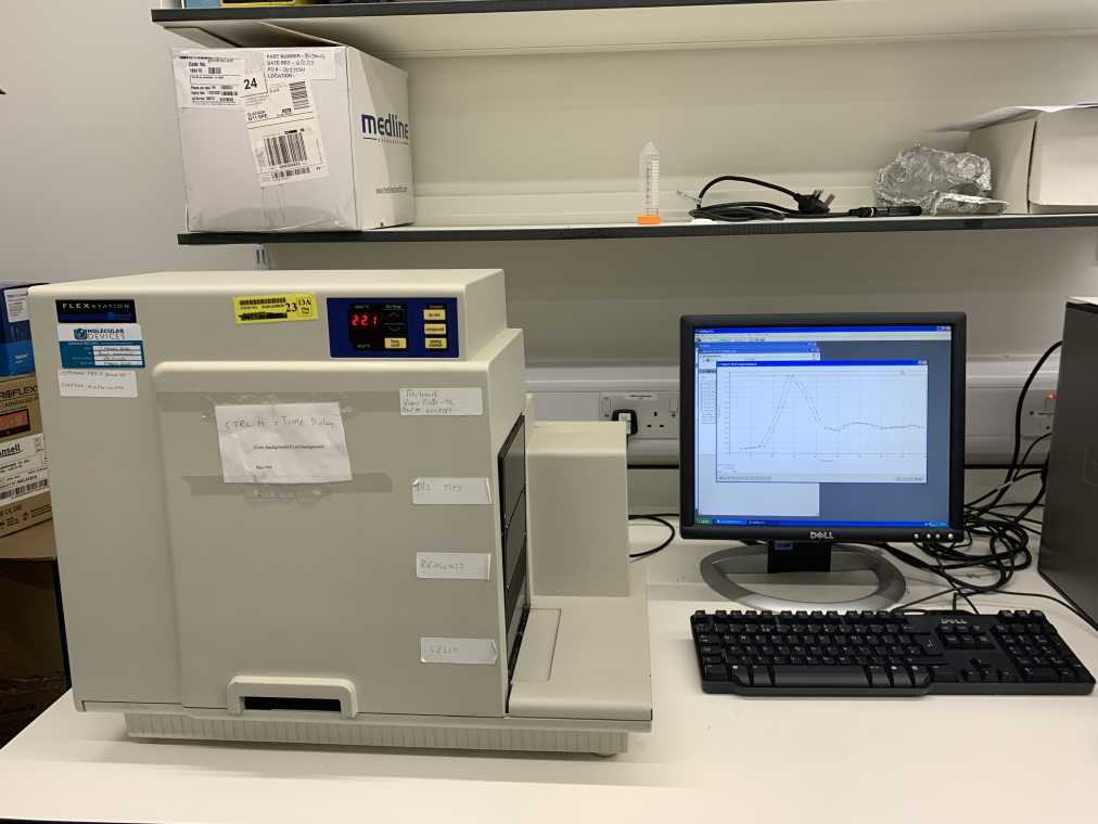
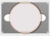
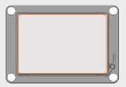
Flexstation II
The Flexstation II instrument has an integrated dual scanning monochromator for optimal multi sequential excitation and detection of assay multi-fluorescent probes. Fast kinetic assays are achieved by using an integrated 8 channel pipettor which can transfer multiple compound fluids very rapidly from the reagents plate to the assay plate and data is acquired as soon as the compound fluid has been dispenced into the well. This property allows fast kinectic events, such as the mobilisation of calcium (Ca2+) from internal stores, extracellular Ca2+ influx into the cytosol via voltage operated Ca2+ channels or changes in membrane potential to be measured in real time.
The integrated dual scanning monochromators can perform excitation or emission spectral scans at 1nm intervals over the excitation wavelength range from 250 to 850 nm and emission wavelength from 375-850 nm. This feature allows users to optimise their assays by determining the actual excitation and emission peak of the fluorescent dye that they are working with. The instrument also has temperature control, automatic shaking and can perform top or bottom read measurements from 6-96 well microplates. Multiple read measurements within a well are also possible when the well scanning mode feature within the software is selected.
The instrument is located within the ARC building on level 4, room 449. To use the instrument contact Dr John Pediani using the email address below:
A picture of the Flexstation II instrument is shown below:
Thermo Fisher Scientific Instruments
Evos FL Auto2 imaging station.
Our invert EVOS FL Auto2 imaging station is a bench top epifluorescence and transmitted light, (bright field/phase contrast and colour) imaging station. It can be automated to tile scan multi-well plates, culture flasks, petri-dishes and slides.
A picture of the epifluorescence imaging instrument is shown below:
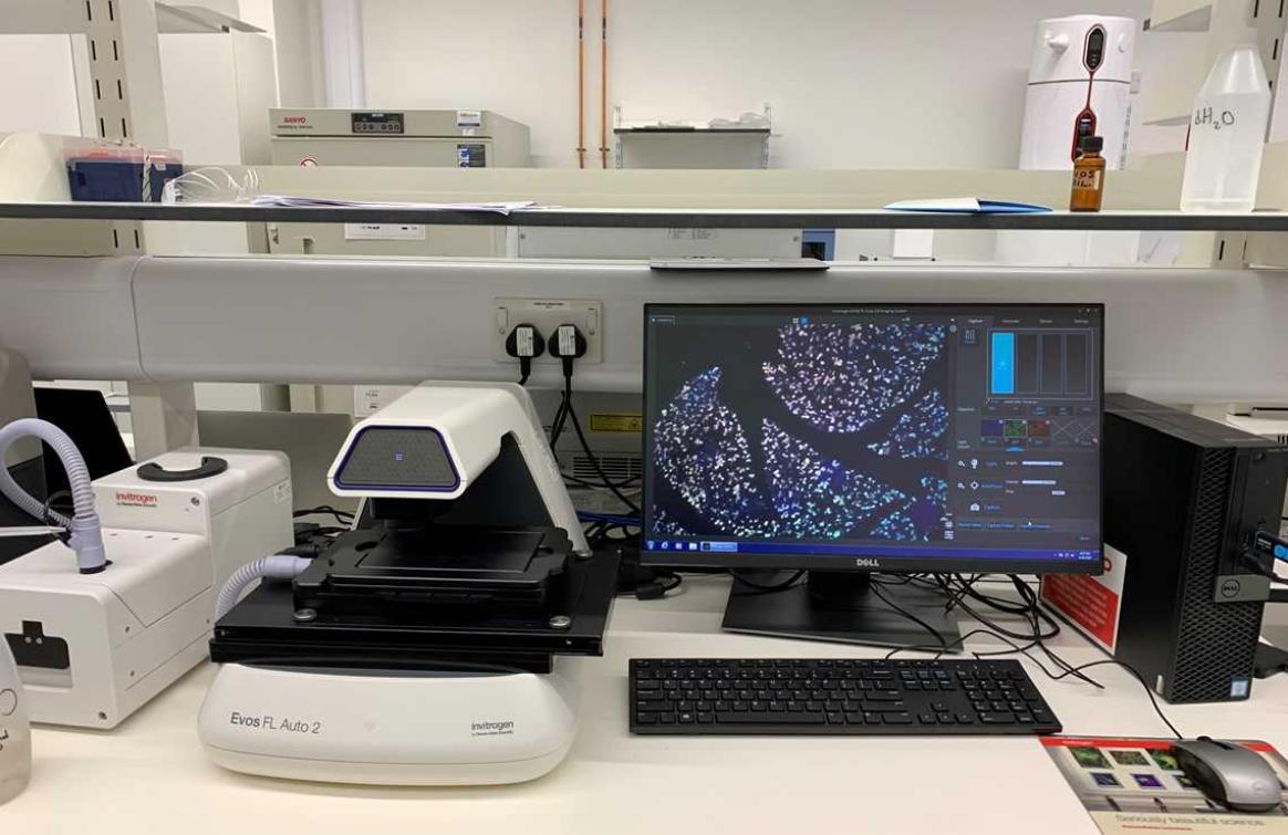
A dedicated monochrome camera, optimised for fluorescence detection, is used to record 16 bit depth multi-fluorescence channel images from live or fixed cell based samples. A separate color camera is also available for recording colorimetic images such as chromogenically stained immunohistochemistry tissue samples. An onstage incubator can also be fitted to perform a wide range of biological assays under physiciological conditions. The system can be fitted with a wide range of LED/filter cubes to sequentially detect fluorescence emitted from dyes over the UV to IR range.
The 3 LED light cubes that we have fitted for sequentially acquiring single plane or z-stack multi-fluorescence channels over time are listed below:
DAPI: Ex = 357/44 nm. Em = 447/60 nm-------- -Cat no: AMEP4650
EGFP: Ex = 470/22 NM. Em = 510/42 NM-----------Cat no: AMEP4651
REPI: Ex = 531/40 nm. Em = 593/40 nm-----------Cat no: AMEP4652
This web link shows the full range of LED light cubes that are currently available to purchase and the dyes that can be detected using our fitted LED light cubes.
The instrument is located within the ARC building on level 4, room 449. To use the instrument contact Dr John Pediani using the email address below:
Stage x-y scanning speed:
Sequential 3 color multichannel fluorescence imaging of a 96 well plate takes < 5 minutes.
Fitted Objective Lens:
A diverse range of coverslip corrected objective lenses, (1.25 x to 100 x), can be purchased depending upon your requirements, e.g. long working distance, (WD), with low numerical aperture; (NA) & short WD with high NA. Lenses fitted are listed directly below:
Turret Position 1-cat no AMEP4699:
40 x Plan Fluorite Dry Air, NA = 0.75; WD = 0.89 mm, phase contast NO.
Turret Position 2-cat no AMEP4622:
4 x Plan Fluorite Dry Air, NA = 0.13; WD = 19.7 mm; phase contrast NO.
Turret Position 3-cat no AMEP4734:
20 x Plan Apochromat Dry Air, NA = 0.75; wd = 0.77 MM; phase contrast NO.
Turret Position 4-cat no AMEP4682:
20 x Plan Fluorite Dry Air, NA = 0.4; WD = 3.1 mm; phase contrast YES.
Turrent Position 5-cat no AMEP4700:
100 x Plan Fluorite Oil immersion, NA = 1.28; WD = 3.1 mm; phase contrast NO.
Other available objectives:
cat no AMEP4682:
2 x Plan Achromat Dry Air, NA =0.06; wd = 5.1 MM; phase contrast NO.
cat no AMEP4681:
10 x Plan Fluorite Dry Air, NA= 0.25; WD = 9.2 mm; phase contrast YES.
cat no AMEP4753:
10 x Plan Apochromat Dry Air, NA = 0.4; WD = 3.27 mm; phase contrast NO.
Stage plates and vessel holders
We have a diverse range of stage plates and vessel holders which can be fitted to suit your sampled needs and these are listed below.
Available Stage Plates
Master plate:
-cat no AMEP-VH035: master plate for the EVOS onstage incubator, accommodates environmental chamber. L = 185.3 mm, W = 135.3 mm.
Slides:
-cat no AMEP-VH021: holds 2 microscope slides or chamber slides with retention clip. L = 76.7 mm, W = 26.4 mm.
-cat no AMEP-VH067: holds 4 microscope slides with retention clip. L = 77 mm, W = 26 mm.
Well plates/well dishes:
-cat no AMEP-VH004: holds one Nunc 100 mm petri dish, D= 88.5 mm.
-cat no AMEP-VH022: holds one multiple with retention clip for AMEPVH001 through to AMEPVH018. L = 128.2 mm, W = 86.2 mm.
Vessel Holders:
Petri dish:
-cat no AMEP-VH029: onstage incubator, holds one 35 mm petri dish with retention clip, diameter = 41 mm.
Well plate:
-cat no AMEP-VH028: onstage incubator, holds one multi well plate with retention clip, L=128.2 mm, W = 86.2 mm.
Features:
- Automated time lapse imaging including area scanning and multifield.
- Z stack visualisation
- Movie AVI creation
- Transmitted light: bright field/phase contrast/color and fluorescence