

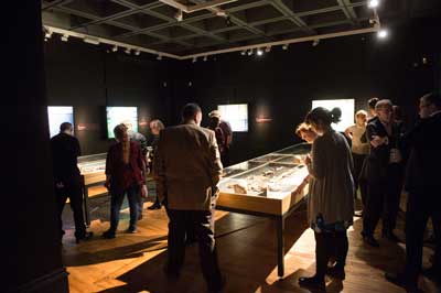
Skeletons: Our Buried Bones
19 August 2016 - 8 January 2017
Hunterian Art Gallery
Admission free
Introduction
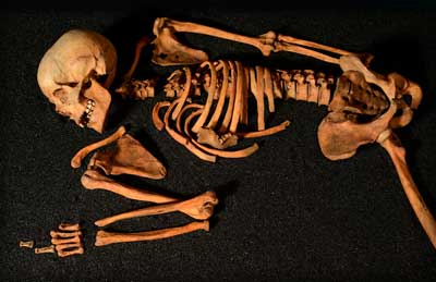
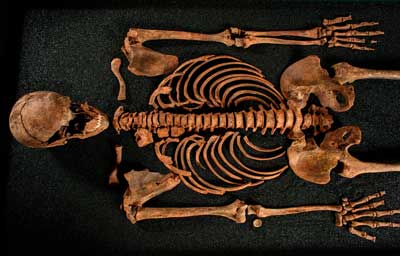
 This special exhibition unearths the stories behind four skeletons from the Museum of London’s 20,000-strong collection and four from burial grounds across Scotland. On display together for the first time, they reveal rich and eventful life stories, with individuals coming from diverse locations and periods of time, from the plague pits of urban London to the beaches of South Uist.
Using the latest scientific methods, careful analysis by experts has provided insights into the health and history of each individual, bringing to life stories that have long been hidden beneath the ground. Each skeleton exhibits pathologies that expose the multiple challenges of life in the past, from fractures and trauma, cancer and the effects of syphilis, to rickets, arthritis and tooth decay.
Research has also shed new light on the grounds they were discovered in and the circumstances in which they were buried.
Skeletons: Our Buried Bones is a collaboration between Wellcome Collection and the Museum of London, touring to Glasgow, Bristol and Leeds over 2016-2018.
Balevullin, Tiree (Neolithic)
 In 1912 a small group of burials were discovered on the machair at Balevullin. Records show four skeletons were found, including a young child aged between 4 and 7 years old. They were located in the vicinity of an Early Iron Age settlement and it seemed likely the burials were of a similar date. However, recent stable isotype analysis revealed that the individuals lived in the Neolithic period. This establishes Balevullin as a rare example of a Neolithic inhumation cemetery. Notes made by A. Henderson Bishop, a wealthy Scottish businessman and pig farmer who led the excavation, describe how one of the skeletons was ‘collared’ by the Professor of Anatomy at the University of Glasgow for his museum. Bishop also states that the excavation led to ‘trouble with the Islanders who accused me of digging up their ancestors and taking them to London to put in a museum’. The other skeletons remained on the Islands and were likely reburied by members of the local community. The skeleton which entered the Anatomy Museum at the University of Glasgow was later transferred to the Hunterian Museum, within whose collections it remains today.
The distinctive shape of the sternum of this individual would have led to a ‘pigeon-chested’ appearance in life. The deformation of the bone could have been a result of childhood rickets, caused by vitamin D deficiency. This would make this the earliest case ever recorded. However, it may also be a congenital condition caused by a disruption in the development of the sternum. The sacrum (tailbone) is fused to the lower lumbar vertebra, a condition known as sacralisation. These changes, along with asymmetry observed in the skull, could suggest a fault along the midline of the body.
It is not always straightforward to identify the sex of skeletal remains. This individual was previously identified as a probable female, but recent analysis suggests that this might have been a possible male.
Extensive stable isotype testing conducted by the University of Bradford revealed that despite the island location this individual didn’t eat fish but a diet of plants and protein from land.
Collection: The Hunterian, University of Glasgow.
Image: © Callum Bennetts - Maverick Photo Agency.
Horse Cross, Perth (Medieval)
 Archaeological excavations were undertaken in Perth in 2003 in advance of the construction of a new concert hall. A small population of burials associated with the medieval chapel of St Laurence were revealed. Archeo-magnetic dating techniques on a hearth sealed below the floor of the church established a date of 1360-95CE. A grave was found a short distance from the main cemetery, near to where the Perth Museum and Art Gallery stands today. The nature of this outlying burial and unhealed injuries found on the skull suggest this may not have been a legitimate burial, but a victim of violence concealed in a shallow grave.
The site was previously home to a royal castle, which was destroyed by flood in 1209. The area was then given to the Blackfriars monastery, and developed into an industrial suburb. Later activity on the site included a series of clay-lined tanning pits and the 17th century horse market which gives the area its name.
This is the skeleton of a teenager, most likely male. The epiphyses – the rounded ends of the long bones – are not yet fused to the shafts, meaning that they could have continued to grow. Despite his youth there is evidence that he worked hard during his life. The marked muscle attachments suggest heavy strain, possibly caused by manual labour. Periostitis, a healed infection of the tissue covering the tibia, can be caused by muscle overuse and is a common cause of shin splints.
The slight curvature of the spine (scoliosis) could be congenital – present from birth. The bowing in the lower legs suggest childhood rickets, caused by vitamin D deficiency, which was common to the urban poor.
The injury to his skull may have occurred at the time of his death. His body was concealed in an outlying grave from the main cemetery burials. This suggests foul play, possibly even murder. However peri-mortem injuries (those that occur at the time of death) are very difficult to distinguish in archaeological remains.
Collection: Perth City Museum and Art Gallery
Image: © Callum Bennetts - Maverick Photo Agency.
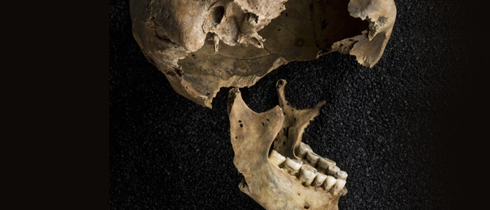
East Smithfield Black Death (1348-1350)
 In the 14th century a virulent strain of the plague arrived in London to devastating effect. It is thought that the outbreak, which struck in 1348, wiped out between a third and half of London’s population. The numbers of the dead were so large that special ‘catastrophe’ burial sites were created to accommodate them. The site at East Smithfield was the first dedicated Black Death cemetery in London. Excavations revealed bodies buried five deep, with children placed between adults to maximise the space. Although buried in mass graves, they were neatly stacked and properly arranged according to Christian tradition, with their heads at the west and feet to the east. After the plague subsided the catastrophe sites were closed, and the Cistercian abbey of St Mary of Grace was built on the East Smithfield grounds. The site was later home to the Victualing-Office and the Royal Mint.
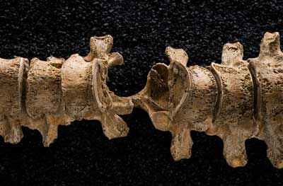
This is the skeleton of a medieval male, thought to be aged 36-45 years. It was recovered from one of London’s ‘catastrophe’ sites created to accommodate victims of the plague in the 14th century. This individual was found with an iron projectile — most likely an arrowhead — lodged in his spine. The bone surrounding it has healed, indicating he recovered from the attack and lived with the object in situ for some time.
Collection: Museum of London.
Image: Courtesy of the Museum of London/Wellcome Images.
Cross Bones (1598 – 1853)
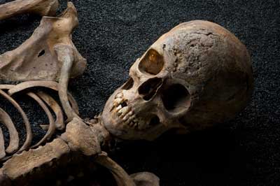 The infamous Cross Bones burial ground served the poor of the parish of St Saviour’s in Southwark. Originally established in the 17th century as a cemetery for ‘single women’ – a euphemism for prostitutes – it had, by 1769, become a paupers’ graveyard. The site lay on unconsecrated ground and was described by John Stow in his Survey of London (1598) as having been made ‘far from the Parish church’. By 1853, after 200 years of use, the ground was finally closed by order of the council. A report published the previous year stated that ‘it is crowded with dead and many fragments of undecayed bones, some even entire, are mixed up with the earth of the mounds over the graves’. Skeletons exhumed from the site in 1992 revealed conditions associated with disadvantaged circumstances, such as rickets and syphilis.
Excavated from the Cross Bones cemetery for paupers and prostitutes, this skeleton shows evidence of two conditions associated with London’s poor. The scarring on the skull is from the ulcererated lesions caused by syphilis. The distinctive bowing of the long bones in the legs indicates rickets; a disease which was common in 19th century London following the rapid industrialisation of the city. This individual is a post-medieval female, thought to be aged between 18 and 25 years old.
Collection: Museum of London.
Image: Courtesy of the Museum of London/Wellcome Images.
Events
Skeletons 10 Minute Talk Programme
Every Wednesday from 28 September - 14 December 2016
1.00pm
Hunterian Art Gallery
Admission free
28 September 2016
Pox and Prejudice? The emotional history of syphilis
Mona O’Brien, Hunterian Associate
5 October 2016
Radiocarbon Dating
Dr Philippa Ascough, Scottish Universities Environmental Research Centre
12 October 2016
Isotopes: Reconstructing life histories through analytical science
Dr Philippa Ascough, Scottish Universities Environmental Research Centre
19 October 2016
Bones, isotopes and ice cores, and the long road to unleaded fuel
Dr Beverly Bergman, Institute of Health and Wellbeing, University of Glasgow
26 October 2016
Make or Break: Visualising bone biology, development and disease
Hussain Jaffery, Hunterian Associate
2 November 2016
Neolithic Skeleton from Balevullin, Tiree: A complex and controversial case!
Dr Stuart McDonald, School of Life Sciences, University of Glasgow
9 November 2016
Medieval Skeleton from Horsecross, Perth: A murdered teenager?
Dr Stuart McDonald, School of Life Sciences, University of Glasgow
16 November 2016
No talk this week
23 November 2016
No talk this week
30 November 2016
Pictish Burial: Pagan or Christian?
Dr Adrián Maldonado, School of Humanities, University of Glasgow
7 December 2016
Chronological Chromosomes: Uncovering DNA through time
Frances Osis, Hunterian MUSE
14 December 2016
Morbid Curiosities: The Ethics of Displaying Human Remains
Taryn Gouck, Archaeology/Celtic Civilizations student, University of Glasgow
Skeletons Meet the Expert
Thursdays 6 October, 3 November and 1 December 2016
5.30pm
Anatomy Museum
Admission free - booking required
Inspired by the Skeletons: Our Buried Bones exhibition, this event offers an outstanding opportunity to meet two leading experts from the University of Glasgow's Anatomy Facility. Three dates are available. Dr Stuart McDonald will begin with a tour of the Anatomy Museum from a historical perspective, followed by Professor Fabio Quondamatteo talking about the 'Skeleton and Locomotor System.' Please note that this will take place in the Anatomy Museum in the Thomson Building. Book your place via Eventbrite.
From Trowels to Test Tubes: Reconstructing ancient human lives with isotope science
Saturday 5 November 2016
10.00am - 1.00pm
Hunterian Art Gallery
£15.00 - booking required
Join Dr Phillipa Ascough to find out about reconstructing ancient human lives with isotope science. This half day event in partnership with the Centre for Open studies aims to introduce the role of isotope science to understand past human lives in archaeology; explain the basics of how isotope techniques work for skeleton analysis and provide practical experience in applying isotope data to archaeological samples. Participants will enjoy a lecture to introduce the concept of isotope science and its application in archaeology; a practical demonstration; a tour and practical exercises in the Skeletons exhibition in the Hunterian Art Gallery and a discussion of findings and interpretations.
Acknowledgements
 Skeletons: Our Buried Bones is a collaboration between Wellcome Collection and the Museum of London, touring to Glasgow, Bristol and Leeds over 2016-2018.