

Advancing 7T Neuroimaging with an In-House, Novel Head Coil
Published: 13 January 2020
Advancing 7T Neuroimaging with an In-House, Novel Head Coil
At the RF coil lab in the Imaging Centre of Excellence, University of Glasgow, we have developed a new dual-mode head coil which supports both imaging modes with a single device and provides a significant cost saving to users of 7T MRI equipment.
In contrast to the current commercial option, the new coil provides whole-brain coverage (Figure 1). This extended anatomical detail will facilitate integration of 7T MRI into clinical pathways and enable whole-brain studies in neuroscience. In addition, the antenna design in the new head coil was tailored to allow for two large cut-outs. This makes the setup less claustrophobic and improves patient comfort and supervision.
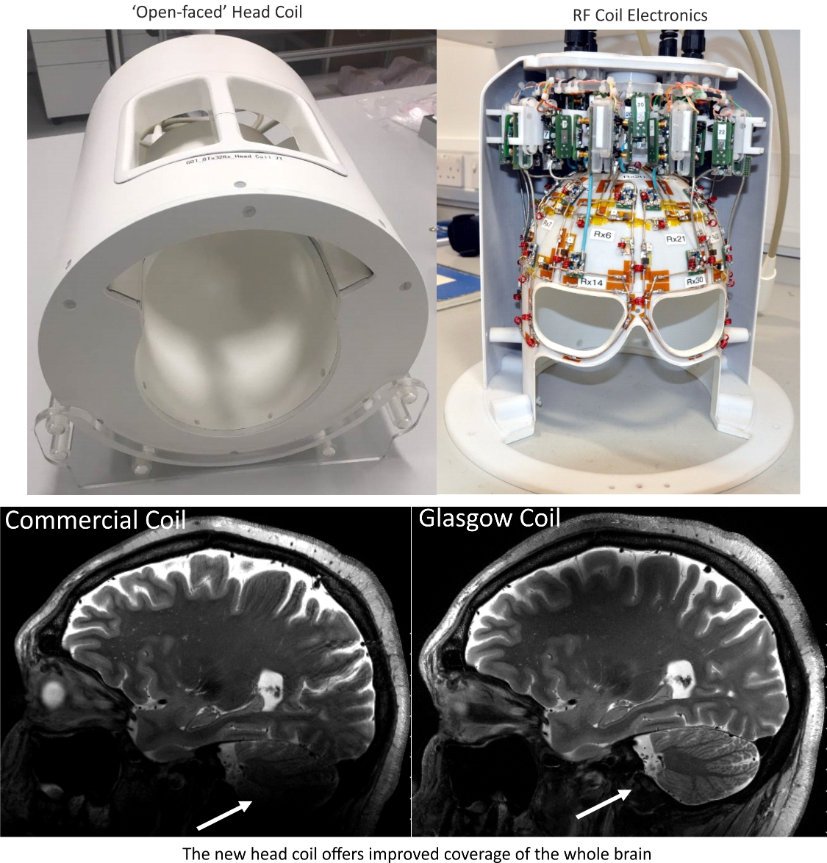
(Figure 1: Comparison between the standard commercial coil and the new Glasgow dual-mode coil)
Magnetic resonance imaging at a magnetic-field strength of 7 tesla (7T) provides increased anatomical detail compared to conventional lower-field MRI at 1.5T and 3T. However, radiofrequency (RF) coil design at this higher field strength is challenging because of the shorter wavelength in tissue compared to conventional MRI. This results in an inhomogeneous image that restricts whole-brain-imaging applications in neuroscience and diagnostic radiology.
This problem can be addressed by using parallel transmit (pTx) MRI scanners together with multi-channel-transmit RF coil arrays. However, pTx technology is still under development and routine scanning at 7T is still performed in a single-channel (sTx) mode. Commercial MRI scanners at 7T support both modes of operation, but require a separate RF head coil for each of the two imaging modes. This makes it cumbersome to switch between the modes and results in a considerable additional expense.
For the second half of 2019 a project team consisting of ICE researchers from the INP, NHS, and Siemens Healthcare have worked to experimentally validate and approve the safety of the Glasgow coil for operation in pTx mode. This requires a more advanced, model-based system for managing real-time power deposition than with sTx operation, for which the coil was previously approved in April 2019. In November, the Clinical Research Imaging Forum Scientific Assessment Group approved use of the coil for pTx applications, and an initial study cohort of 10 healthy volunteers has begun.
Figure 2 shows results from the first volunteer using the pTx imaging mode compared to images acquired in the sTx mode. These preliminary results already demonstrate an improvement in the field homogeneity and image quality in the pTx case.
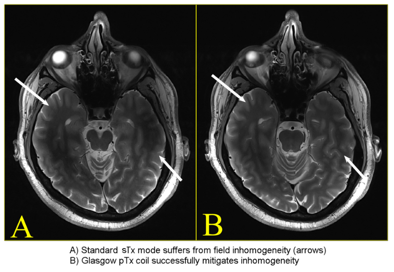
Figure 2. Comparison between Glasgow coil in conventional sTx (top A) and pTx (bottom B) modes
Project Team: Sydney N. Williams, Sarah Allwood-Spiers, Paul McElhinney, Yuehui Tao, Gavin Paterson, John E. Foster, David A. Porter, Shajan Gunamony
First published: 13 January 2020