

WWCRC Microscope Facility
The Institute of Cancer Sciences microscopy facility is based at the Wolfson Wohl Cancer Research Centre (WWCRC) on the Garscube Campus of the University of Glasgow. We have three types of microscope which are available for communal use within the Institute. All our communal microscopes are from ZEISS and the associated software is very user friendly.
Basic Axio Vert microscopes are supplied in all the tissue culture rooms and enable convenient acquisition of low quality images. For better quality images we also have the Axio Observer microscope and the LSM 780. The LSM 780 is specialised for capturing high resolution fluorescent images. The Axio Observer can be used for widefield fluorescence microscopy or for transmitted light microscopy and has been adapted for live cell imaging.
The facility is operated on a not-for-profit basis. The charges for using the equipment cover the costs of servicing, maintaining, and updating the equipment.
University staff and students out with the Institute may use our microscopy facility. Please ask the facility manager about availability.
Microscopy facility manager:
Mr Allan McVie
Telephone: 0141 330 8124
Microscopes
ZEISS Axio Vert A1
Located in the cell culture suites (Rooms 115, 116, 217, 218, 317, 318, 417, and 418) in the WWCRC
 Booking: not required
Charge: not applicable
Light sources: LEDs
Filter sets: GFP and mRFP
Objectives:
- X5
- X10
- X20
- X40
Other features:
- Phase condenser
- Motorised stage
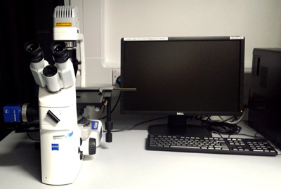
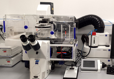
ZEISS Axio Observer A2
Located: room 123 in the WWCRC
Booking: http://bookings.tcrc.gla.ac.uk/equipment
Charge: £15/ hour during core hours (9am – 5pm) except for Live Cell Imaging (LCI) warm up time which is £30 for 4 hours (you will need to make a note of this in your booking), LCI O/N (5pm – 9am) is £7.50/ hour.
 Light sources:
- Halogen (HAL) bulb for transmitted light microscopy
- Mercury-Xenon (HXP) bulb for widefield fluorescent microscopy
Filter sets:
- DAPI (49), ex BP 365, em BP 445/50
- GFP (38 HE), ex BP 470/40, em BP 525/50
- DsRed (43 HE), ex BP 550/25, em BP 605/70
- Cy 5 (50), ex 640/30, em 690/50
Objectives (application):
- X2.5 EC Epiplan Neofluar (brightfield/ fluorescence)
- X5 EC Epiplan Neofluar (brightfield/ fluorescence)
- X10 EC Plan Neofluar (brightfield/ DIC/ fluorescence)
- X20 LD Plan Neofluar (brightfield/ phase contrast/ DIC)
- X40 Plan Neofluar (brightfield/ phase contrast/ DIC/ fluorescence)
- X40 Plan Neofluar (brightfield/ phase contrast/ fluorescence)
- X63 Plan Apochromat (brightfield/ DIC/ fluorescence)
- X100 Plan Neofluar (brightfield/ DIC/ fluorescence)
Other features:
- Phase and DIC condensers
- Adapted for live cell imaging with temperature and CO2 control within an incubator
Applications:
- Advanced tile scans
- Time lapse
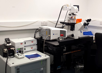
ZEISS LSM 780 Confocal Microscope
Located: room 123 in the WWCRC
Booking: http://bookings.tcrc.gla.ac.uk/equipment
Charge: £30/ hour
 Light sources:
- Halogen (HAL) bulb for transmitted light microscopy
- Laser lines for confocal microscopy (405, 488, 514, 561, 633)
Objectives (application):
- X10 EC Plan Neofluar (brightfield/ phase contrast/ fluorescence)
- X20 Plan Apochromat (brightfield/ DIC/ fluorescence)
- X40 EC Plan Neofluar (brightfield/ DIC/ fluorescence)
- X63 Plan Apochromat (brightfield/ DIC/ fluorescence)
Other features:
- Floating table
- Motorised stage
Applications:
- Tile scans
- Z stacks
- Co-localisation analysis
- Spectral unmixing of fluorochromes with overlapping excitation and emission spectra
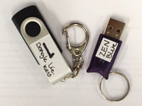
Analysis software dongle
We have a ZEN blue (and ZEN black) software dongle available which enables you to perform analysis and processing of images on your personal computers.
Before using the dongle for the first time, you will need to perform an installation using the CD and licence key.
 Located: room 107 in the WWCRC
Booking: http://bookings.tcrc.gla.ac.uk/equipment
Applications:
- Splitting multi-channel images
- Stitching tiles
- Compressing Z stacks
- Deconvolution
- Quantitative analysis of staining using the Image Analysis wizard
- Co-localisation analysis
Rules for use
• You must book your session for the Axio Observer and the LSM 780 in advance using the online booking system.
• If you need to cancel your session, you must alter your online booking.
• If you cancel your session after the start time of the session you will still be charged for it
• Users must take care to start up and shut down the microscopes in the correct order.
• If no-one is booked on after you, make sure that you completely shut down the microscope
• If someone is booked on after you, you don’t have to turn everything off, but leave the lasers on idle (not run) on the LSM 780.
• You must only use oil on the appropriate objectives (this is indicated on the touch screen display): the X40 and X63 on the LSM 780; the X63 and X100 on the Axio Observer.
• After using oil, please ensure you have cleaned the objective with lens tissue and methanol.
• Other than cleaning objectives, do not perform any other kind of maintenance on the microscope.
• Objectives and other components are only to be changed Jessica – contact her if you have a problem or require components to be changed.
• The Axio Observer is the default microscope for LCI.
• Users must allow for up to 4 hours warm up in their LCI booking.
• A single user cannot book LCI more for more than 3 consecutive days.
Data storage policy
All our microscopes are networked and enable you to login with your GUID and password, and then to save images directly to your personal H: drive.
Where possible, you should avoid saving data on the C: drive and instead save/ transfer data to your personal H: drive.
Periodically, reminders will be circulated for users to remove their data from the microscope computers, and if this is not carried in due time, data will be deleted.
Safety
Laser safety: When acquiring images with the LSM 780 and the lasers are on, you must not insert any object in the path of the laser and avoid looking directly at the laser beam.
Sharps: A sharps bin is provided for the disposal of any broken slides or coverslips.
Biological safety: If you accidentally spill some media when performing live cell imaging, you should clean the stage with 70% ethanol and notify Jessica. The microscopy facility is a Containment Level 1 facility and therefore you cannot undertake work requiring Containment Level 2. Before imaging any samples originating from a Containment Level 2 tissue culture room you will need to perform a risk assessment for the work and the Biosafety or GM committee must then approve that this work represents no or negligible risk (Class 1).
For further information on safety, please contact:
Stacey Hoare
Safety co-ordinator
email Stacey.Hoare@glasgow.ac.uk
telephone 0141 330 8707
Liz Musgrove
GM safety officer
email Liz.Musgrove@glasgow.ac.uk
telephone 0141 330 7282
Carol McCormick
Biological safety officer
email Carol.McCormick@glasgow.ac.uk
telephone 0141 330 3509
Guidelines for Image Publication
Different journals will have different requirements regarding the formatting of images for publication and therefore it is best practice to refer to the guidelines issued by the relevant journal. Look at the links below for some general information on standards of imaging for publication.
Nature Publishing Group - Image Integrity and Standards
http://www.nature.com/authors/policies/image.html
(accessed 16/03/2016)
Journal of Cell Biology - Image Acquisition
http://jcb.rupress.org/content/172/1/9.full.pdf+html
(accessed 16/03/2016)
NCBI – Ethical Guidelines for Image Manipulation
http://www.ncbi.nlm.nih.gov/pmc/articles/PMC4114110/pdf/nihms575182.pdf
(accessed 16/03/2016)
Links
The Carl Zeiss Microscopy Online Campus
http://zeiss-campus.magnet.fsu.edu/index.html
The Olympus Microscopy Resource Centre
http://olympus.magnet.fsu.edu/index.html
Nikon Microscopy Education
BD Spectraviewer
http://www.bdbiosciences.com/eu/s/spectrumviewer
IHC world
http://www.histology-world.com/
DAKO IHC
Thermo Fisher IHC
Royal Microscopy Society