

Abstracts
Poster 1: Abdullah Akaydin
SilverGuardian: A Privacy-Preserving Framework for In-Home Human Activity Recognition and Gait Assessment for Health Monitoring
Abdullah Akaydin* (University of Glasgow), Julien Le Kernec (University of Glasgow), Aleksandra Vuckovic (University of Glasgow), Haobo Li (University of Dundee), Jon Clarke (GJUNH), and Swati Chopra (GJUNH)
Human activity recognition (HAR), gait assessment, and fall detection play vital roles in health monitoring by enabling both timely intervention and early disease prevention. Radar sensing has emerged as a competitive solution compared to conventional approaches such as cameras, wearable devices, or other RF-based techniques, offering unobtrusive operation, robustness to environmental conditions, and preservation of privacy. Leveraging these advantages, radar-based HAR and gait analysis provide valuable opportunities for the early detection and continuous monitoring of diverse health conditions. Neurological disorders such as Parkinson’s disease, Alzheimer’s disease, and stroke can be inferred from gait abnormalities and irregular activity routines. Musculoskeletal disorders, including arthritis and osteoporosis, manifest as reduced mobility and impaired posture transitions, while cardiovascular and pulmonary diseases are reflected in diminished activity levels and interrupted movement patterns. Furthermore, activity monitoring supports fall risk assessment and provides indicators of mental health conditions such as depression. By integrating these capabilities, the proposed framework establishes a foundation for future radar-enabled health monitoring systems aimed at supporting prevention, timely intervention, and long-term well-being.
Poster 2: Alicia Gardiner
Alicia Gardiner, Research Assistant, James Watt School of Engineering
On the Cutting Edge - FEA simulation of ultrasonic bone surgery
Ultrasonic bone cutting tools show significant promise in surgical applications for reducing post-operative complications in patients, such as: potential infection, neurovascular injury and osteonecrosis. A novel miniaturized ultrasonic surgical cutter prototype is presented as a comparative case study, involving ex-vivo testing on porcine cortical rib bone, to validate a cutting-edge FEA model. Key parameters investigated in these studies is frictional-based temperature increase and user applied cutting force. Recording, and predicting, cutting force and temperature can be applied to recommend ideal operation of the device to use it safely and minimize post-surgical complications.
A competitive feature of this FEA model is its reduced computational requirements and high processing speeds for 30+ seconds of explicit/dynamic simulated time, which enables unparalleled comparison of experimental datasets over longer durations. Additionally, the model incorporates a natural sawing cadence to better reproduce experimental conditions, which is generally not incorporated in typical machine tooling or crack propagation methods applied currently to simulate ultrasonic cutting. These simulation methodologies are a powerful predictor of device performance, with potential to expedite the design and testing of ultrasonic surgical cutters, assisting in their transition into industrial applications – particularly robot-mounted cutters in Da-Vinci Surgical Systems.
Poster 3: Callum Macaulay
Callum Macaulay, University of Glasgow - Statistics, PhD student
Accelerating Patient-Specific Biomechanical Model Calibration with Surrogate Optimisation
Patient-specific biomechanical models are powerful tools for clinical decision-making, but their practical use is often hampered by the time and computational cost of calibrating them to an individual's unique biology. This paper proposes using surrogate optimisation to overcome this challenge. Instead of repeatedly running computationally intensive simulations, we create a computationally cheap surrogate model (such as a Neural Network or a Gaussian Process) that learns the relationship between a model's parameters and its output. This allows us to rapidly and efficiently find the best parameters for a specific patient's data.
This approach is particularly relevant for time-critical clinical applications. For instance, when planning cardiovascular interventions, accurately modelling a patient's arterial stiffness or compliance is essential for predicting outcomes and assessing risk. Our method has the potential to dramatically reduce the time needed to calibrate these models, bringing us closer to real-time, patient-specific simulations. This will not only improve the accuracy of predictions but also enable robust uncertainty quantification, giving clinicians a more complete picture of a patient’s unique biomechanics and helping to improve treatment planning.
Poster 4: Chia-Yin Wu
Improving transmit magnetic field homogeneity using a neurovascular head and neck coil with tailored parallel transmission pulses for MRI at 7T
Chia-Yin Wu (1), Divya Baskaran (1), Keith Muir (1), Natasha E. Fullerton (1,2), Shajan Gunamony (1,3), David A. Porter (1)
1 Imaging Centre of Excellence, University of Glasgow, Glasgow, UK
2 Department of Neuroradiology, Institute of Neuroscience, NHS Greater Glasgow and Clyde, Glasgow, UK
3 MR CoilTech Limited, Glasgow, UK
Abstract:
A neurovascular head-and-neck coil (NVHN) [1] provides coverage of the brain and cervical spine enabling neurovascular MRI techniques a 7T. 7T MRI systems improve signal-to-noise ratio and enables faster imaging but capabilities are challenged by non-uniform transmit magnetic fields (B1+). RF pulse design using parallel transmission (pTx) efficiently mitigates B1 + non-uniformity. With a dedicated NVHN coil, RF pulse designs can remain streamlined without the need of coordinating multiple coils imaging different anatomical regions. We demonstrate initial insight using a NVHN coil with tailored pTx pulses to improve signal uniformity across the brain and neck region in vivo.
Scans were performed on a 7T Terra Siemens system using a custom-built 8-transcieve-56-receive NVHN coil. Standard circularly polarised pulses were compared with tailored pTx pulses [2,3,4] to evaluate excitation fidelity across the whole brain and upper neck region using the 3D gradient-recalled echo sequence.
Significant signal uniformity improvements were achieved in the cerebellum, peripheral regions of the head and central neck region in pTx-mode. A median of 22% improvement in excitation fidelity was achieved using pTx pulses across five subjects. This shows promising potential in incorporating pTx pulses in more advanced MRI techniques with targeted interest in both the brain and neck regions.
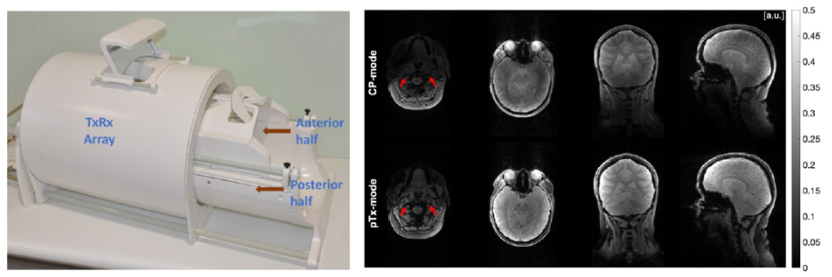
Figure. Left: 8TxRx56Rx Neurovascular Head-and-Neck (NVHN) coil (Image from [1]). Right: T2*-weighted images acquired with standard pulses in circularly polarised (CP-mode) and with tailored pTx pulses (pTxmode)
Acknowledgements: This project is funded by Scottish Funding Council, grant H07012/242700324, the Christine Rodgers endowment fund, the Neuroscience Foundation, the Strength in Places fund, and Innovate UK through The Living Laboratory: driving economic growth in Glasgow through real world implementation of precision medicine (award Ref: 107140). The authors also thank Robin Sayer and Iain Hendry from the NHS Medical Devices Unit for providing the mechanical design for the NVHN coil.
References
[1] D. Baskaran, B. Ding, S. Chu et al. Magn Res Med. 70(1): 386-400 (2024). [2] M. A. Cloos et al. Magn Res Med. 67(1): 72-80 (2012). [3] W. Grissom, C.-Y. Yip, Z. Zhang et al. Magn Res Med. 56(3): 620-629 (2006). [4] K. Setsompop, L. L. Wald, V. Alagappan et al. Magn Res Med. 59(4): 908-915 (2008).
Contact: Chia-Yin Wu, Postdoctoral Researcher
Imaging Centre of Excellence, University of Glasgow | e-mail: Chia-Yin.Wu@glasgow.ac.uk
Poster 5: Janhavi Ghosalkar
T1 mapping with multi-contrast MP-RAGE and 2D GRAPPA at 7T MRI
Janhavi Ghosalkar (1), Graeme A. Keith (1), Belinda Ding (1,2), Natasha Fullerton (3), Shajan Gunamony (1,4), David Porter (1)
(1) Imaging Centre of Excellence, University of Glasgow, Glasgow, Scotland (2) NHS University Hospital Birmingham,
Birmingham, UK (3) NHS Greater Glasgow and Clyde, Glasgow, UK (4) MR CoilTech Limited, Glasgow, UK
Abstract:
High-resolution T1 -weighted imaging at 7T benefits from MP2RAGE’s [1,2] reduced sensitivity to inhomogeneities that affect image quality at ultra-high-field MRI. This study extends MP2RAGE by acquiring multiple inversion times (TIs) using a modified multi-contrast MPRAGE (McMPRAGE) sequence with Cartesian readout and 2D GRAPPA [3] acceleration, enabling efficient sampling for quantitative T1 mapping.
Scans were performed on a 7T MRI system using a custom head coil [4], acquiring phantom and in vivo data at 0.86mm isotropic resolution. T1 estimation was performed using the Deichmann-Haase correction [5] and an updated model accounting for incomplete relaxation during the delay time (TD). Monte Carlo simulations showed that in comparison to Deichmann-Haase correction that underestimates T1, the updated model improves accuracy, and precision under certain fitting conditions.
McMPRAGE produced contrast comparable to MP2RAGE and shows promise for enabling robust T1 estimation across TDs without significantly increasing scan time. Improved T1 mapping can enhance tissue characterisation in neurological disorders such as multiple sclerosis and Alzheimer’s disease. Future work will apply the updated model to in vivo data and explore integration with transmit field mapping and inversion efficiency (IE) estimation to improve precision in quantitative imaging.
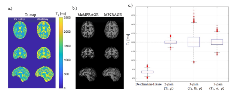
Figure: a.) T1 maps in three planes for two different TDs with a GRAPPA acceleration factor of 3x2. b.) Acquired MP2RAGE compared to a ‘calculated’ MP2RAGE generated using estimated signal values extrapolated from voxel-wise T1 fitting. c.) Boxplot showing the results of a 10,000-sample Monte Carlo simulation for two different model functions. The two and three-parameter fittings use the same updated model function where the Alpha symbol is the flip angle and the Rho Symbol is the product of spin densities and receiver sensitivities. The dotted line at 1200ms depicts true T1.
References:
[1] Mugler JP, Brookeman JR. Magn Res Med. 15(1):152-157 (1990). [2] Marques JP, Kober T, Krueger G, van der Zwaag W, Van de Moortele PF, Gruetter R. Neuroimage. 49(2):1271-1281 (2010). [3] Griswold MA, Jakob PM, Heidemann RM, et al. Magn Res Med 47(6):1202-1210. (2002). [4] Williams SN, Allwood-Spiers S, McElhinney P, et al. Frontiers in Physics. (2021). [5] Deichmann R, Haase A. Jou of Magn Res. 96(3):608-612 (1992).
Contact:
Janhavi Ghosalkar, PhD student
Imaging Centre of Excellence, University of Glasgow
Email: j.ghosalkar.1@research.gla.ac.uk
Poster 6: Joe Knapper
Joe Knapper, Department of Physics, Research Associate
Early diagnosis is a key factor in improving patient outcomes, yet access to reliable diagnostic hardware and trained users remains limited in many regions. Optical microscopy is the "gold standard" for the diagnosis of a wide range of conditions globally, including malaria and many cancers. We present the OpenFlexure Microscope, a locally manufacturable, robotic microscope that uses 3D-printed parts and off-the-shelf components. The microscope incorporates automated sample scanning, sub-micron mechanical precision, and digital imaging capabilities suitable for integration with pathology lab workflows, medical education, and machine learning pipelines. As an open-source device, the OpenFlexure Microscope has been reproduced in over 60 countries, including local manufacturing and deployment in hospitals in Rwanda, Brazil, and the Philippines. These initiatives have enabled digitisation of histological biopsies and cytology smears in regions with limited access to digital pathology tools. This has facilitated new opportunities for pathologists to present at international seminars, while medical students gain access to digitised slides that represent their future clinical practice. We highlight the technical achievements of this work, including sample scans and details of our public educational seminars in global pathology, as well as the social impact of enabling diverse regions to produce and maintain their own medical devices.
Poster 7: Kaso Osman
Authors:
- Dr. Kaso Osman (GMC: 7998877) BSc (Hons) Pharmaceutical and Medicinal Chemistry, MBChB (University of St Andrews & University of Dundee, 2023) - completed foundational training August 2025.
Current role: Locum resident Doctor / Senior House Officer (SHO) - Dr. Paul McNamara (GMC: 7134256) BSc (Hons) 1st class Anatomy, MBChB (University of Glasgow, 2011) MRCGP Hons & Distinction
Title
Artificial Intelligence in Clinical Practice: Perceptions of Healthcare Professionals and Medical Students
Background
Artificial Intelligence (AI) is gaining prominence in healthcare. While its potential to aid diagnosis and decision-making is widely acknowledged, perspectives of its integration remain insufficiently explored.
Objective
To assess perceptions of AI among healthcare professionals and medical students, focusing on benefits, risks, ethics, and anticipated changes to practice.
Methods
A cross-sectional online survey (May 1–7, 2025) targeted healthcare professionals. Domains included AI exposure, confidence in integration, and training needs. Demographic and professional data supported subgroup analysis.
Results
168 professionals responded, 61% had >10 years of experience and 60% reported prior AI use. Most (62%) viewed AI as supportive rather than replacement. Confidence was limited: 42% trusted current diagnostic abilities, but only 8% believed AI could independently manage major clinical decisions. Anticipated adoption was highest in Radiology (89%), Pathology (63%), and Dermatology (40%). Just 1% felt adequately trained; 46% stressed the need to supervise and interpret AI outputs. Barriers included ethical/legal concerns (65%). Additionally, 43% feared AI could reduce training opportunities.
Conclusions
Findings show cautious optimism for AI, highlighting the need for adoption that supports, rather than replaces, clinical expertise.
Keywords
Artificial Intelligence, Emergency Medicine, Radiology, Clinical Practice
Poster 8: Marija Vaskeviciute
Marija Vaskeviciute, School of Physics & Astronomy, Research Assistant
Aortic stenosis is the most prevalent valvular heart disease in ageing populations, affecting nearly 8% of individuals over 84 years in Western countries. Early diagnosis is critical to improving treatment outcomes, yet existing portable diagnostic methods often require lengthy acquisition times and multiple chest sensor placements, limiting their utility in real-world settings. We report preliminary findings from an early-stage clinical validation study of a novel, lightweight laser-camera device that remotely detects mechanical vibrations related to valve motion from a single chest location. Our approach achieves over 90% diagnostic accuracy for aortic stenosis with only a 10-second recording time. These results demonstrate the potential of laser-based vibrometry as a rapid, non-contact digital health tool for accessible and scalable valvular disease screening.
Poster 9: Said Al Multlq
Multiome profiling of the uromodulin knock-out mouse
Al Multlq S, Zhou M, Graham D, McBride MW
School of Cardiovascular and Metabolic Health, University of Glasgow, Glasgow, UK
Introduction: Primary hypertension, a major cause of cardiovascular morbidity and is tightly linked to renal function. Genome-wide association studies have significantly associated uromodulin (UMOD) promoter variants to hypertension. To define UMOD’s functional role, we applied multi-omics profiling to the Umod knock out mouse under basal and high-salt conditions.
Methods: Wild-type (WT) and Umod knock out (KO) mice were fed a normal diet or 2% salt in their drinking water (n=3 per group). A multi-omics approach combined bulk RNA sequencing (padj<0.5), proteome (FC>1.2), metabolomics (p<0.05) analyses and interpreted using Ingenuity Pathway Analysis.
Results: Bulk RNAseq identified UMOD-dependent transcriptional shifts in mitochondrial function, ER stress, unfolded protein response, and cilium assembly. Proteomic and metabolomic integration highlighted salt loading suppressed oxidative phosphorylation in WT but enhanced compensatory mitochondrial activity in Umod KO kidneys, indicating genotype-specific, salt-dependent bioenergetic adaptation.
Conclusions: These findings reveal UMOD as a key regulator of renal transcriptional networks and mitochondrial function, with its absence reshaping cellular stress responses and altering adaptation to salt loading. The identified pathways and sodium transport provide mechanistic insight into salt sensitive blood pressure regulation and highlight potential molecular targets for precision strategies to reduce cardiovascular risk.
Poster 10: Shaun Chuah
Title
ChatIBD: AI Companion for Inflammatory Bowel Disease (IBD) Clinicians
Abstract
Inflammatory bowel disease (IBD), including Crohn’s disease and ulcerative colitis, is a growing global health challenge. Managing IBD is increasingly complex, with rapidly evolving treatment options, proliferating guidelines, and varying regional practices. Clinicians face limited time to remain current, and access to IBD expertise is uneven worldwide.
ChatIBD (www.chatibd.com) is a new artificial intelligence (AI) tool designed to support healthcare professionals delivering IBD care. By combining retrieval-augmented generation with curated guidelines, ChatIBD provides clear, evidence-based answers to practical clinical questions, always referenced to trusted sources and tailored to the user’s region. A “deep research” mode extends searches to emerging literature, ensuring clinicians can keep pace with innovation.
Safety and accuracy are reinforced through an integrated dosing database built from European Medicines Agency (EMA) product information, delivering regulator-approved induction and maintenance regimens. ChatIBD’s multilingual capability (English and Spanish in testing) extends accessibility across diverse settings.
Currently in beta testing with over 20 consultant gastroenterologists, ChatIBD has demonstrated >95% accuracy across 400 reviewed responses. By making specialist-level knowledge accessible, ChatIBD reduces unwarranted variation in care, empowers clinicians, and supports better outcomes for people with IBD - while offering a scalable digital health model that can extend to other disease areas/specialties.
Name
Shaun Chuah (shaun.chuah@glasgow.ac.uk)
Department
School of Infection and Immunity
Job Title
Clinical Senior Research Fellow/Honorary Consultant Gastroenterologist
Poster 11: Yazan Haidar
Three-dimensional (3D) printing is transforming orthopaedic practice by bridging imaging, planning, and hands-on rehearsal. Through the NHS Graduate Innovation in Health Technology fellowship, which aims to establish in house clinical 3D printing services in hospitals, two complementary applications are presented: presurgical anatomy visualization and surgical simulation for planning and training. Patient specific models are generated from CT datasets using validated segmentation workflows and printed in rigid bone matching material. For complex deformities, fracture patterns, and tumour resections, tactile models enhance spatial understanding, enable pre-contouring of fixation hardware, and support shared decision making with patients. Parallel simulation models reproduce critical steps like osteotomies, fixation strategies, and implant positioning, allowing teams to test approaches and anticipate challenges. This case study demonstrates an in-house service to create patient specific anatomy for presurgical procedure simulation. The 3D-printed workflow integrates with existing imaging pipelines and governance processes, emphasizing reproducibility, model traceability, and cost-effectiveness at scale. Collectively, these models provide a low-risk, high-fidelity environment to optimize strategy before the first incision, translating preoperative insight into intraoperative efficiency and improved educational outcomes. This highlights a scalable, standards-driven pathway to embed 3D printing across orthopaedic care pathways for measurable clinical and training impact.
Poster 12: Zeqi Luo
General movement assessment (GMA) is a non-invasive diagnostic tool for the early detection of brain dysfunction through the qualitative assessment of general movements, and the development of automated methods can broaden its application. However, mainstream pose-based automated GMA methods are prone to uncertainty due to limited high-quality data and noisy pose estimation, hindering clinical reliability without reliable uncertainty measures. In this work, we introduce UDF-GMA which explicitly models epistemic uncertainty in model parameters and aleatoric uncertainty from data noise for pose-based automated GMA. UDF-GMA effectively disentangles uncertainties by directly modelling aleatoric uncertainty and estimating epistemic uncertainty through Bayesian approximation. We further propose fusing these uncertainties with the embedded motion representation to enhance class separation. Extensive experiments on the Pmi-GMA benchmark dataset demonstrate the effectiveness and generalisability of the proposed approach in predicting poor repertoire movement pattern.