

The Great Escape: How Influenza A Uses Cellular Tunnels to Evade Immune Detection
Published: 16 June 2025
Influenza viruses use secret cellular tunnels to escape your immune system. New research uncovers influenza's most elusive escape route - tunnelling nanotubes that let viruses spread completely undetected.
Influenza A viruses (IAVs) are always on the move. Every year, they stage seasonal outbreaks—and occasionally, pandemics—wreaking havoc on public health. At the core of their success is their ability to invade and spread between the cells of an infected individual.
Once inside, the battle begins—a race between the virus and the immune system. The virus, hijacks cells, forcing them to churn out copies in rapid succession. Our body, sensing this hostile takeover, mounts a defense aiming to neutralize any virus breaking free.
But here’s the twist: imagine the virus as a bank robber, cornered by the authorities that is our immune response. Logic would say it’s trapped—but just like in the movies, there’s always a way out. Even when surrounded, IAVs don’t need to burst out of the cell to spread. Instead, they can perform their own Great Escape—tunneling between cells, bypassing the immune response. In this process referred to as direct cell to cell spread, the virus finds hidden pathways between neighboring cells, continuing its spread, avoding capture
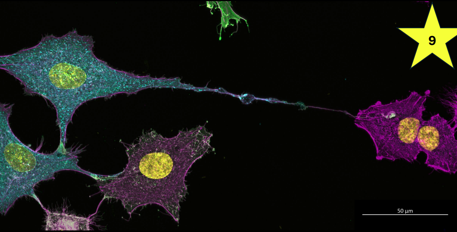
How do they do this? As part of the take over, IAVs don’t just use the machinery of the cell to make more copies, but it uses the cell itself to essentially inject copies of the viral genome into neighbouring cells, initiating a new infection.The virus does this by triggering the cell to produce tunnelling nanotubes (TNTs). TNTs are thin projections of the cell membrane that extend out and touch neighbouring cells, often at impressive distances.This forms a highway for the virus to go on the run and find safe harbour in the newly contacted cell.
Whislt it was known that IAVs could induce the formation of TNTs in the lab, it was not known how they were doing this. Furthermore, we didn’t know if these structures could form in the lungs where IAVs infect, and whether they are used by different IAVs to spread infection.
Using super-resolution fluorescence microscopy, we visualized and quantified TNT-like structures both in cultured cells and within the lungs of infected mice, identifying conditions under which these intercellular connections emerge. Additionally, we used fluorescently labeled viral proteins to determine the efficiency by which different IAVs can spread directly between cells when virus particle release is inhibited with an antiviral drug—providing key insights into non-canonical routes of viral dissemination.
To uncover how IAV infection triggers tunneling nanotube (TNT) formation, we systematically investigated key viral and host factors driving this process. Like piecing together a forensic trail, we tracked the clues IAV leaves behind—examining how viral replication, virion budding, and host cell responses contribute to TNT induction.
For the first time, we observed TNT-like structures emerging from individual cells within the lungs of IAV infected mice. Additionally, even when virus particle release was blocked using an antiviral drug, IAVs still spread efficiently between cells—revealing that alternative pathways for viral dissemination can be readily accessed by these viruses.
Our findings showed that TNT-like structure formation is a common feature of IAV infection but is not triggered by secreted immune signalling molecules or membrane alterations linked to the budding of different virions. Instead, TNT induction relied on active viral replication within the infected cell, combined with the activation of programmed cell death (apoptosis). Interestingly, when we inhibited apoptosis we greatly reduced the ability of IAVs to spread directly between distant cells, indicating that the TNT-like structures triggered by IAV induced cell death could then successfully deliver infection to its neighbours.
Our findings expand our understanding of how direct cell to cell spread contributes to the within-host dissemination of IAVs. By demonstrating the presence of TNT-like structures in IAV infected lungs, we reveal—for the first time—that these structures form not only at infection sites but also within normal lung epithelium.
This discovery suggests that TNTs may play a broader role in both health and disease, emerging even in tissues and organs previously considered challenging environments for fragile nanotube formation. Moreover, our investigation into how IAV triggers TNT-like structures—and the efficiency with which the virus spreads directly between cells—provides deeper insights into the mechanisms shaping infection dynamics.
By understanding how IAV interacts with its host, we can begin to explore strategies to block its spread at the cellular level. Uncovering these covert pathways of viral transmission offers a crucial opportunity to disrupt even the virus’s most elusive movements.
First published: 16 June 2025
<< News