

Skeletons: Our Buried Bones Press Images
Conditions of Use
1. Images must not be masked out, cut down, superimposed with type matter or in any way defaced.
2. The appropriate caption and credit line must be reproduced with the image. All copyright information must be reproduced in full.
 C0046099 Download image
Caption: Skeletons
Credit: Courtesy of the Museum of London/Wellcome Images
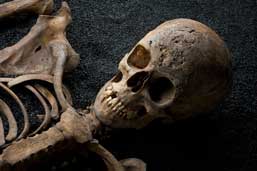
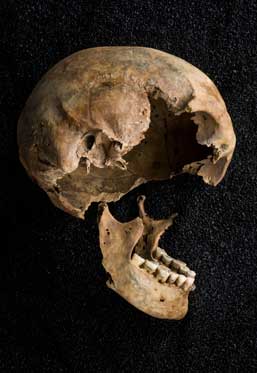
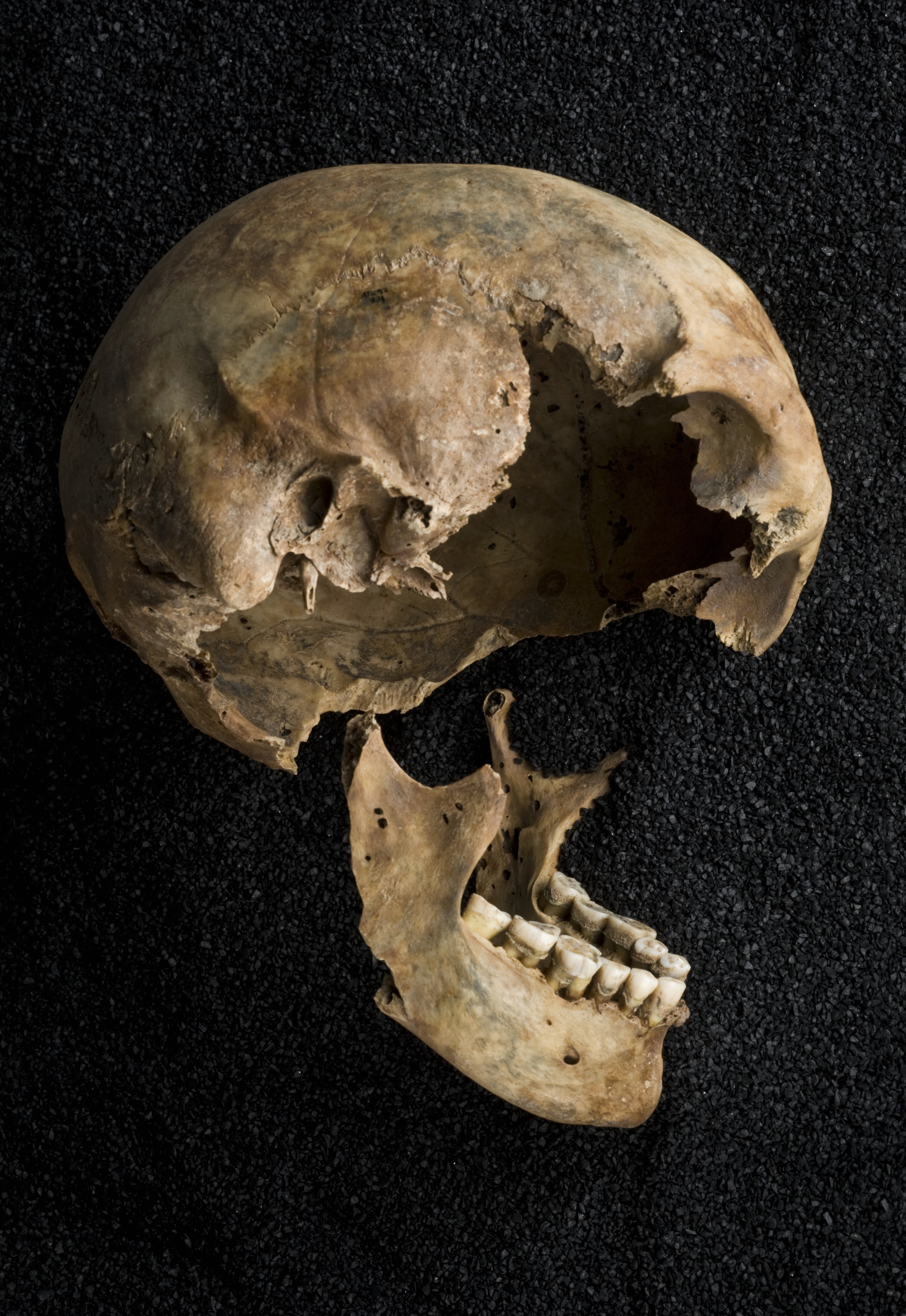
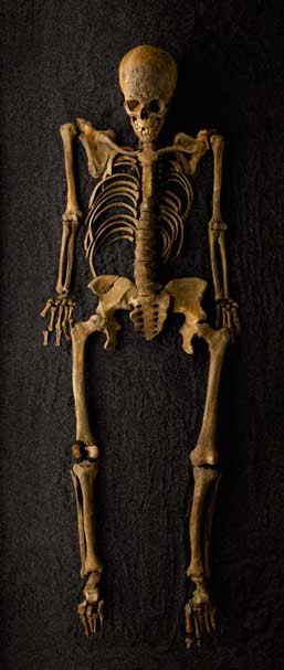
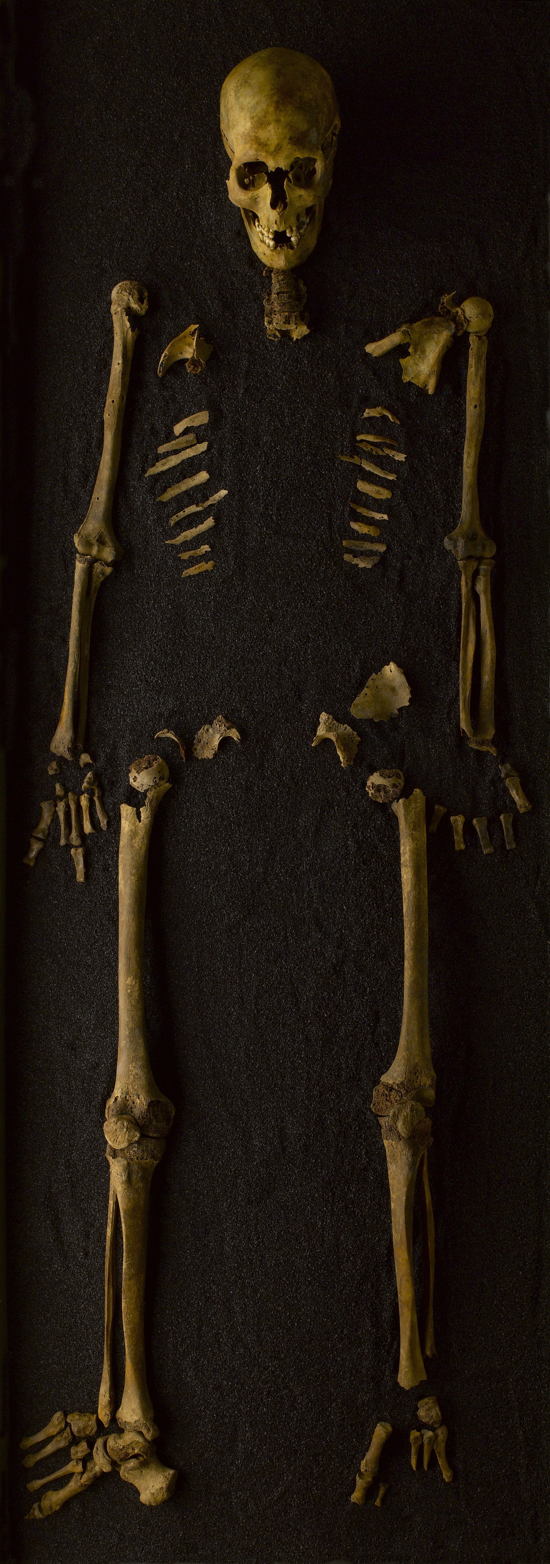
Cross Bones, Redcross Way, SE1 1598 – 1853 / Post-Medieval female / aged 18 – 25
Syphilis, residual residual rickets
Excavated from the Cross Bones cemetery for paupers and prostitutes, this skeleton shows evidence of two conditions associated with London’s poor. The scarring on the skull is from the ulcererated lesions caused by syphilis. The distinctive bowing of the long bones in the legs indicates rickets; a disease which was common in 19th century London following the rapid industrialisation of the city.
 C0046273 Download image
Caption: Skeletons
Credit: Courtesy of the Museum of London/Wellcome Images
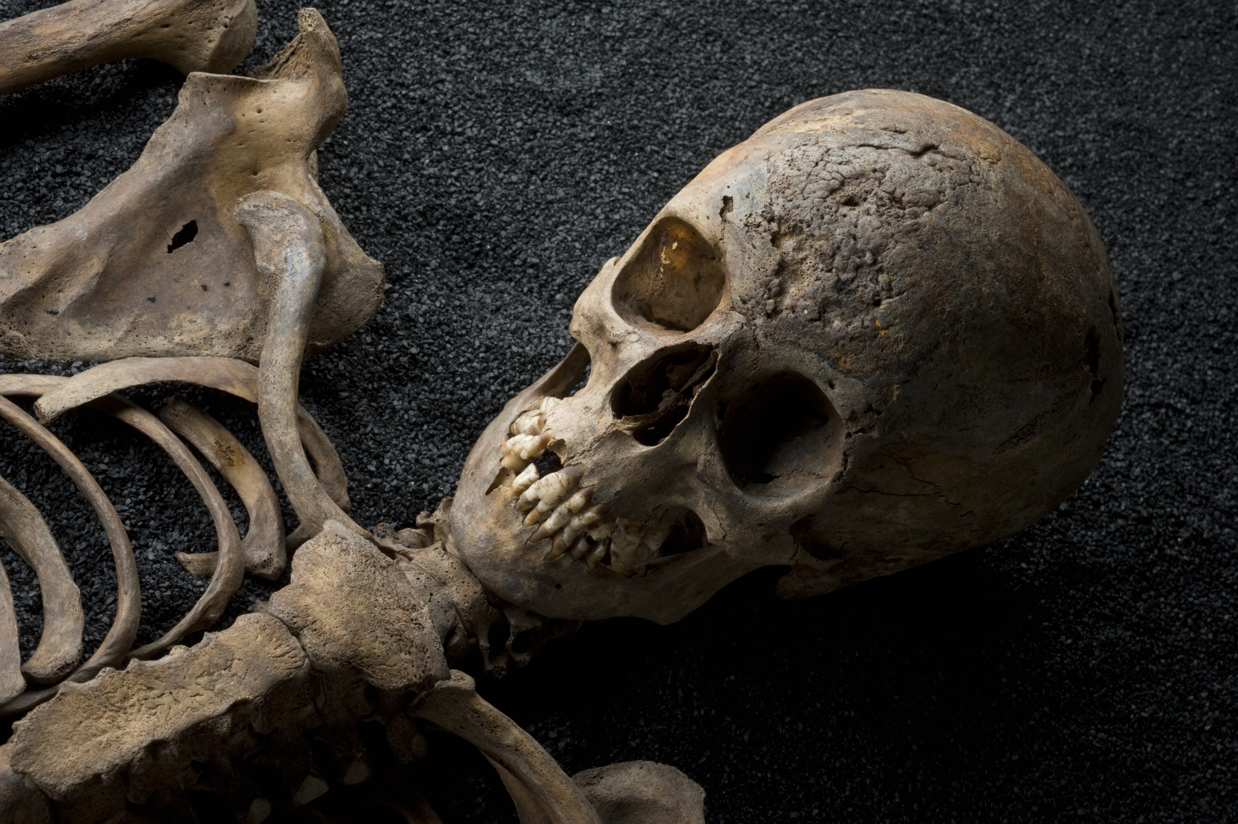
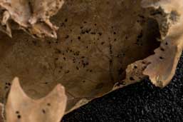
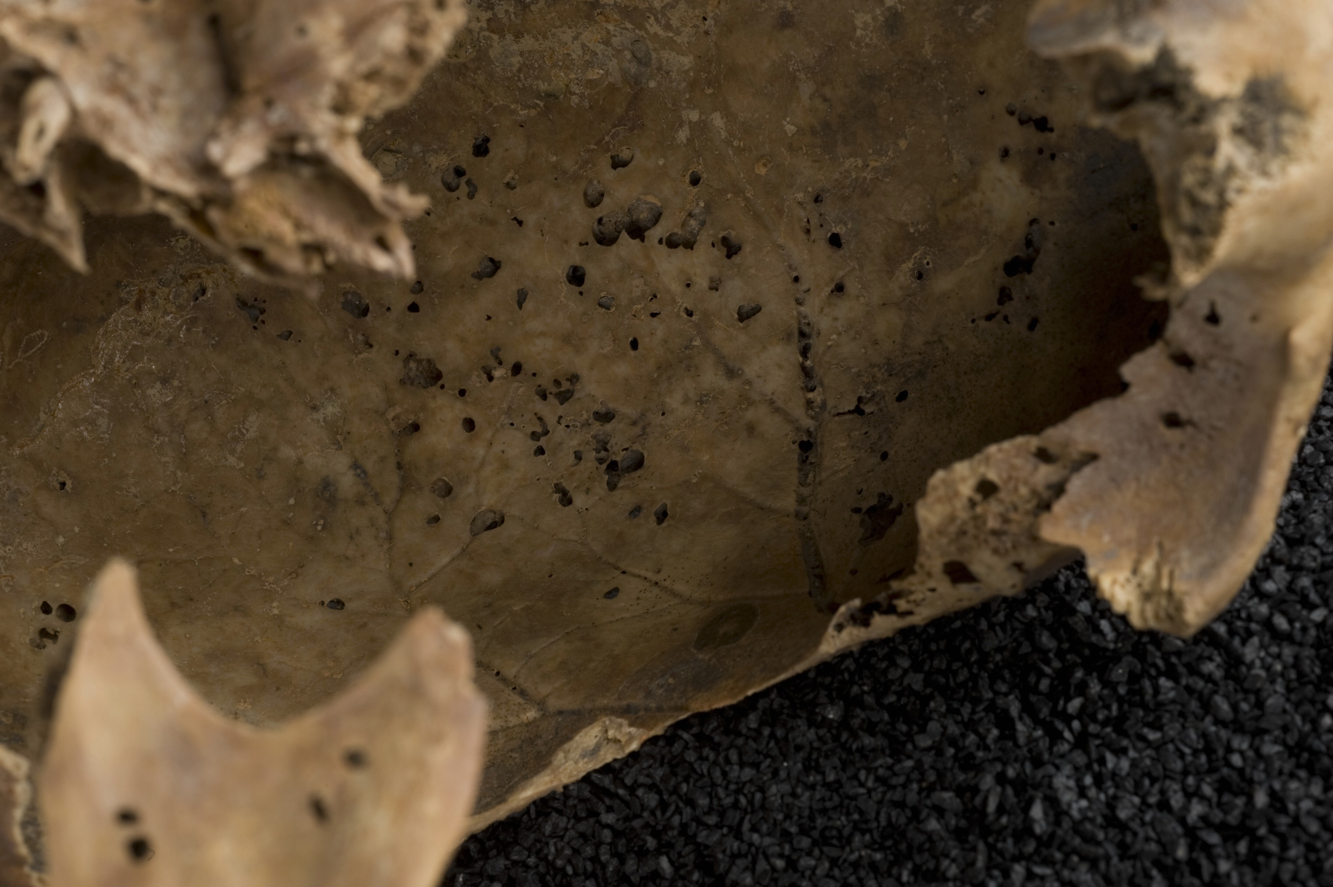
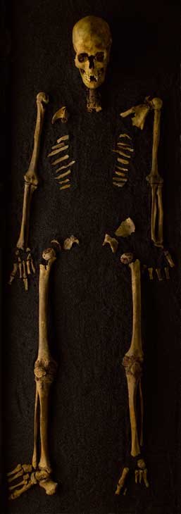
Roman West, 46 – 50 Holborn Viaduct, EC1 1st – 5th centuries / Roman male / age unknown
Small round lesions in skull caused by multiple myeloma
The small holes visible on the jawbone and inside of the skull suggest that this individual was suffering from multiple myeloma — a cancer of the plasma in the blood. This condition causes the bones to soften, creating the lesions. The fissures in the back teeth have been ground flat due to the gritty food that made up the Roman diet. This process helped to protect the teeth from dental caries (decay).
Fractured clavicle (collarbone)
Multiple myeloma also increases production of osteoclasts - substances that normally break down old bones. This causes the bones to weaken further, making fractures more likely. The Roman had a broken clavicle that subsequently healed, causing a slight bump.
 C0046299 Download image
Caption: Skeletons
Credit: Courtesy of the Museum of London/Wellcome Images
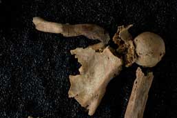
Roman West, 46 – 50 Holborn Viaduct, EC1 1st – 5th centuries / Roman male / age unknown
Small round lesions in skull caused by multiple myeloma
The small holes visible on the jawbone and inside of the skull suggest that this individual was suffering from multiple myeloma — a cancer of the plasma in the blood. This condition causes the bones to soften, creating the lesions. The fissures in the back teeth have been ground flat due to the gritty food that made up the Roman diet. This process helped to protect the teeth from dental (decay).
 C0046303 Download image
Caption: Skeletons
Credit: Courtesy of the Museum of London/Wellcome Images
Roman West, 46 – 50 Holborn Viaduct, EC1 1st – 5th centuries / Roman male / age unknown
Small round lesions in skull caused by multiple myeloma
The small holes visible on the jawbone and inside of the skull suggest that this individual was suffering from multiple myeloma — a cancer of the plasma in the blood. This condition causes the bones to soften, creating the lesions. The fissures in the back teeth have been ground flat due to the gritty food that made up the Roman diet. This process helped to protect the teeth from dental (decay).
Close-up of skull interior
The small holes visible on the endocranium - the inside of the skull - also indicate multiple myeloma. Cancerous cells in the bone marrow inhibit the normal bone-forming cells, causing them to soften and lesions to form.
 C0046356 Download image
Caption: Skeletons
Credit: Courtesy of the Museum of London/Wellcome Images
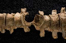
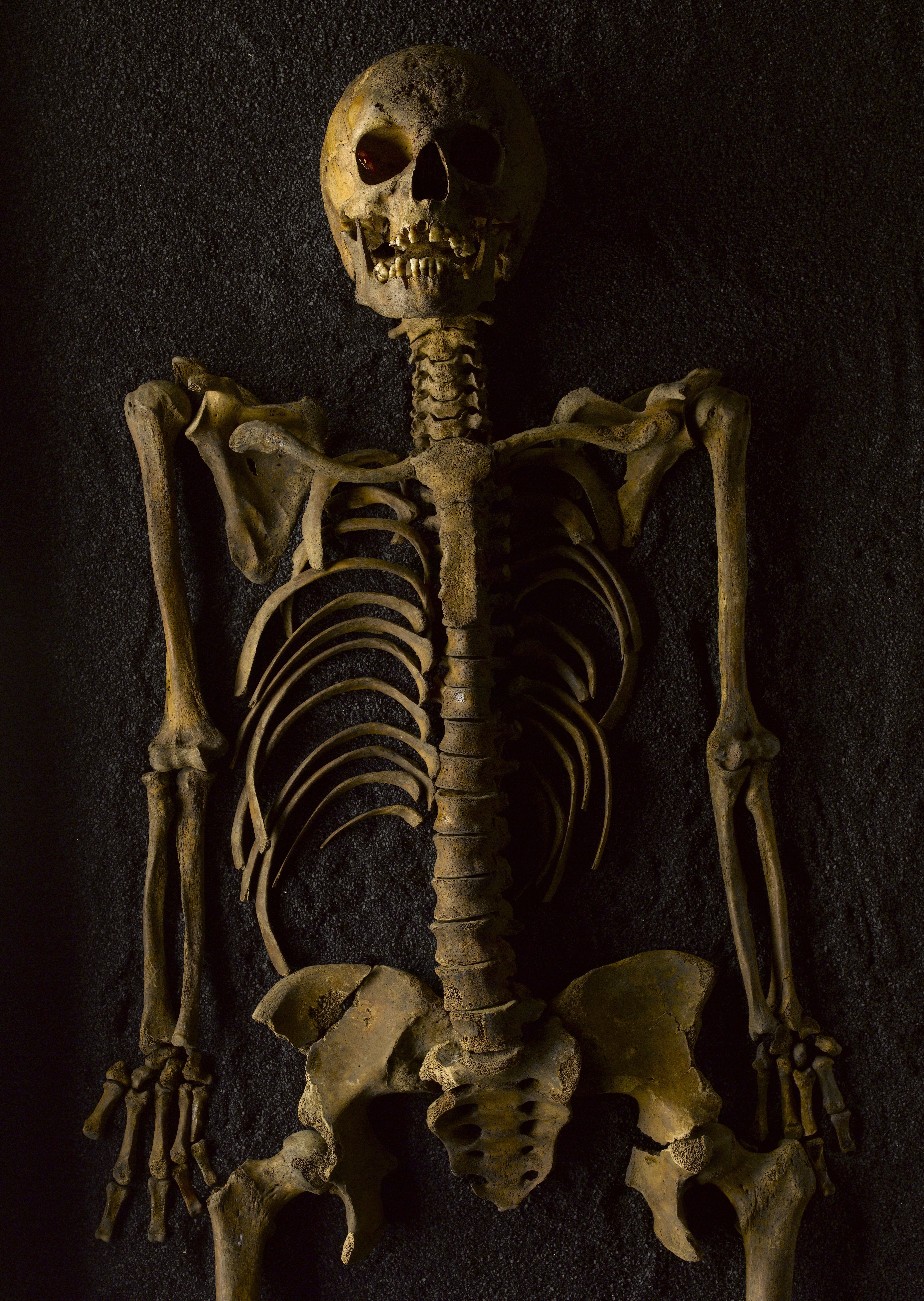
East Smithfield Black Death, East Smithfield, E1 1348 – 1350 / Medieval male / aged 36 – 45 years
Close-up of spine with arrowhead
This individual was found with an iron projectile — most likely an arrowhead — lodged in his spine. The bone surrounding it has healed, indicating he recovered from the attack and lived with the object in situ for some time. The skeleton was recovered from one of London’s ‘catastrophe’ sites created to accommodate victims of the plague in the 14th century.
 C0046427 Download image
Caption: Skeletons
Credit: Wellcome Library, London
Cross Bones, Redcross Way, SE1 1598 – 1853 / Post-Medieval female / aged 18 – 25
Syphilis, residual rickets
Excavated from the Cross Bones cemetery for paupers and prostitutes, this skeleton shows evidence of two conditions associated with London’s poor. The scarring on the skull is from the ulcererated lesions caused by syphilis. The distinctive bowing of the long bones in the legs indicates rickets; a disease which was common in 19th century London following the rapid industrialisation of the city.
 C0046431 Download image
Caption: Skeletons
Credit: Wellcome Library, London
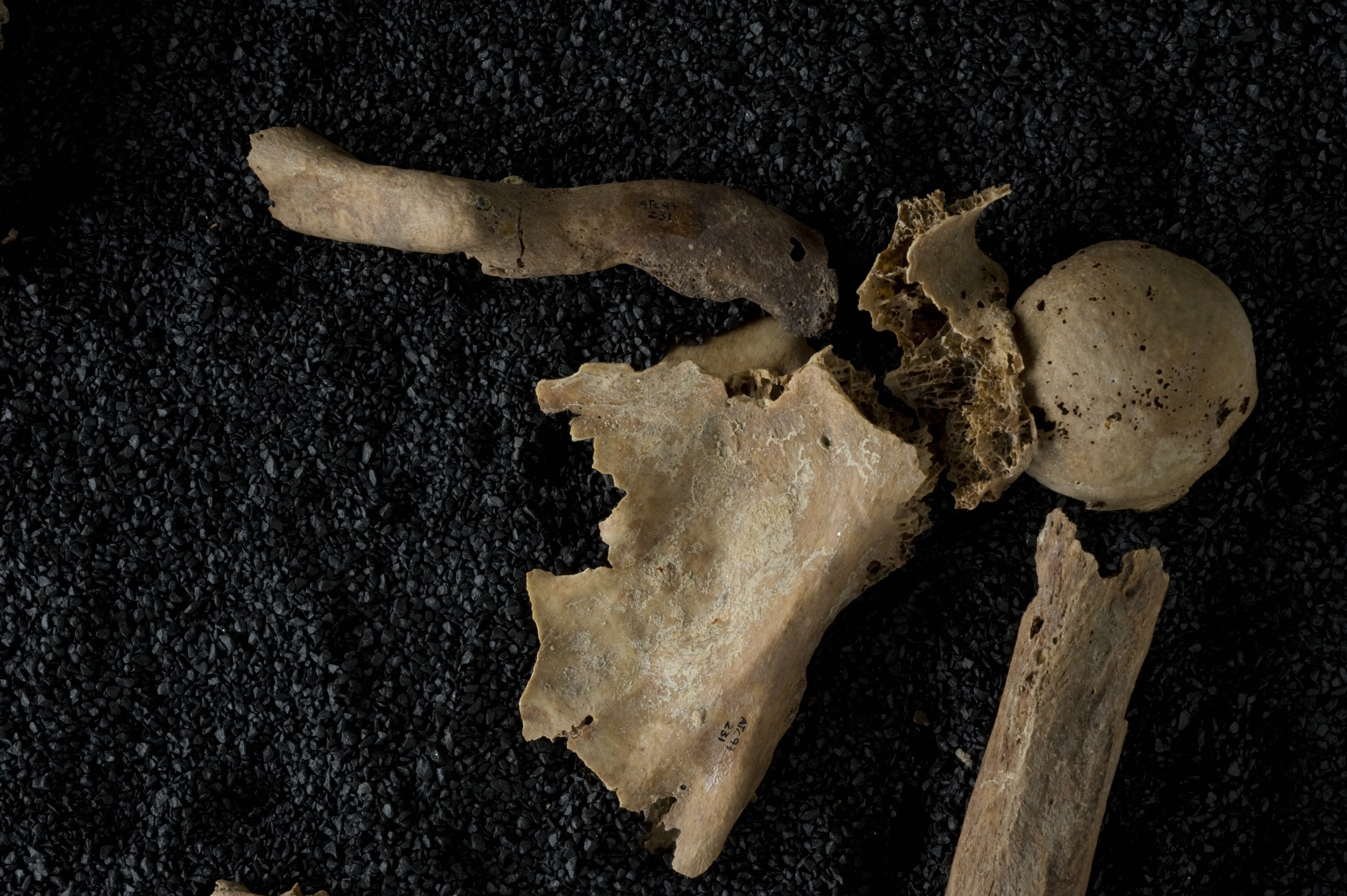
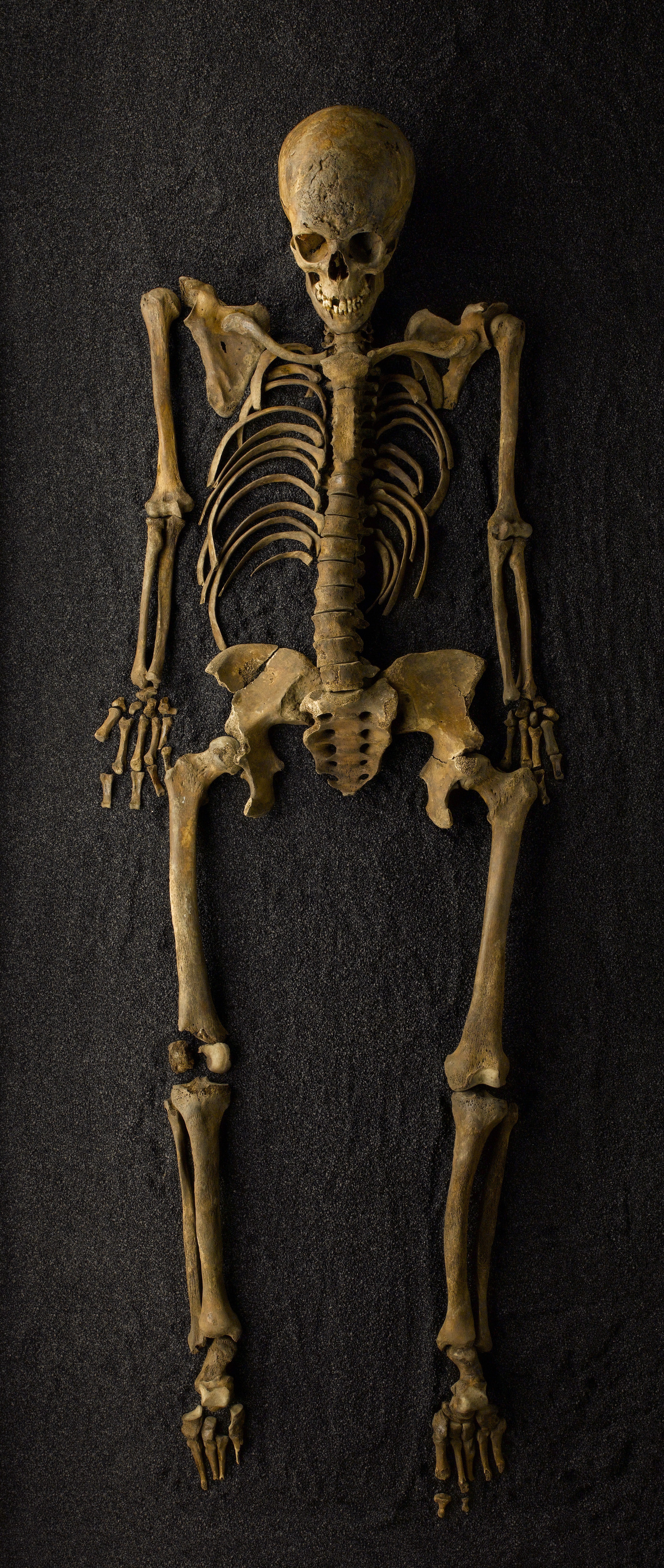
Roman West, 46 – 50 Holborn Viaduct, EC1 1st – 5th centuries / Roman male / age unknown
Small round lesions in skull caused by multiple myeloma
The small holes visible on the jawbone and inside of the skull suggest that this individual was suffering from multiple myeloma — a cancer of the plasma in the blood. This condition causes the bones to soften, creating the lesions. The fissures in the back teeth have been ground flat due to the gritty food that made up the Roman diet. This process helped to protect the teeth from dental (decay).
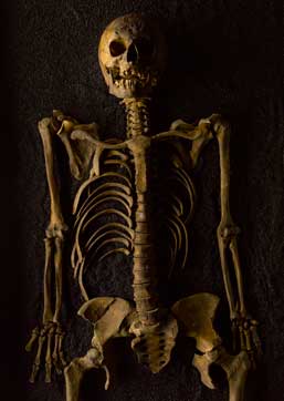
Full skeleton
This skeleton, excavated from the Roman West burial ground, dates from the second to third century CE. It is that of a male of unknown age. The skeleton shows extensive evidence of multiple myeloma - a cancer of the plasma in the blood. This disease weakens the bones and is the likely cause of the fragmentation of the ribs and pelvis.
 C0046434 Download image
Caption: Skeletons
Credit: Wellcome Library, London
Cross Bones, Redcross Way, SE1 1598 – 1853 / Post-Medieval female / aged 18 – 25
Syphilis, residual residual rickets
Excavated from the Cross Bones cemetery for paupers and prostitutes, this skeleton shows evidence of two conditions associated with London’s poor. The scarring on the skull is from the ulcererated lesions caused by syphilis. The distinctive bowing of the long bones in the legs indicates rickets; a disease which was common in 19th century London following the rapid industrialisation of the city.
 L0082078 Download image
Caption: Cross Bones, Thomas Adank, 2008.
Credit: Thomas Adank
 L0082079 Download image
Caption: East Smithfield, Thomas Adank, 2008.
Credit: Thomas Adank
 L0082080 Download image
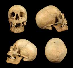
Caption: Human skeleton, Tiree, Scotland, 1912.
Credit: © The Hunterian, University of Glasgow 2016
 L0082081 Download image
Caption: Human skeleton, Tiree, Scotland, 1912.
Credit: © The Hunterian, University of Glasgow 2016
 L0082082 Download image
Caption: Perth, photograph by Thomas Adank, 2016.
Credit: Thomas Adank
 L0082083 Download image
Caption: Roman West, photograph by Thomas Adank, 2008.
Credit: Thomas Adank
 L0082084 Download image
Caption: Skull from human skeleton, Tiree, Scotland.
Credit: © The Hunterian, University of Glasgow 2016
 L0082085 Download image
Caption: Tiree, photograph by Thomas Adank, 2016.
Credit: Thomas Adank