

Functional Materials
 Modern transmission electron microscopes and scanning transmission electron microscopes are allowing us unprecented insights into the nanoworld. The latest generation of microscopes comes with aberration correction for the magnetic lenses used to focus the electrons, which now allows us to produce image resolutions significantly better than atomic size, and to produce electron beams just fractions of an Angstrom across. This allows us to do some fantastic science and this is being used at Glasgow to study the atomic scale structure and chemistry of a wide range of nanostructured materials including:
- Thin film and multilayer oxides for functional applications
- Nanoprecipitates in high manganese steels
- Defects just two atoms wide in doped bismuth ferrite multiferroic ceramics
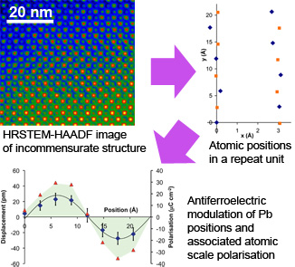
- Novel structures in borderline antiferroelectric lead zirconate titanate (pictured)
- High dielectric constant oxide layers for ultrafast semiconductor devices
Nanoparticle synthesis & characterisation
 Nanoparticles have become a 'big thing' in nanotechnology and can be found in consumer products ranging from suncream to antibacterial socks. Their small size, typically below 50nm in diameter, imbues enhanced and often completely new functionality, including catalytic, magnetic and optical properties.
In collaboration with a number of groups, notably the Murrie group in the School of Chemistry, we have undertaken a number of studies. Highlights include a detailed study of the synthesis of magnetite particles, synthesis of unusual lanthanide nanowires and development of 'octapod' MnO nanoparticles with enhanced magnetic properties that may be suitable for medical applications.
Organic Electronics
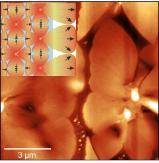 We have recently conducted some scanning probe studies of the nucleation and growth of p-type organic semiconductor molecules within transistor architectures, with the aim to understand how to improve device performance. Further details are available at doi:10.1039/C3TC31783H.
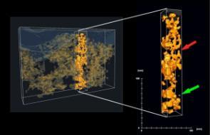
Transmission Electron Microscopy (TEM) tomography volume reconstruction of a hybrid solar cell. The yellow phase represents ZnO nanoparticles forming a network within the polythiophene (P3HT) matrix. The arrows indicate a cul-de-sac (red) and a not connected ZnO particle (see Nature Materials (2009), 8(10), 818-824).