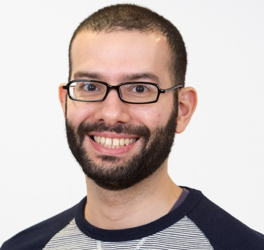
As part of a strategy to integrate imaging technology across the School, an imaging platform for the Sir Graeme Davies Building – named The Glasgow Imaging Facility (GIF) - has been created with Dr Leandro Lemgruber Soares (pictured) appointed as an Imaging Technologist.
Dr Lemgruber, who was previously responsible for the imaging equipment of the Wellcome Centre for Molecular Parasitology, has considerable expertise and experience of light and electron microscopy techniques.
He is your first point of contact for information and support on available techniques and advising on future grants and projects, as well as ensuring access management is simplified and improved.
Our Vision
A centralised imaging platform that will allow the integration of service contracts, attract new users and allow a better integration with partners in other universities and in industry.
Imaging will be the enabling biomedical technology of the next 30 years in the same way that DNA/DNA sequencing has been over the last 30 years.
We anticipate that the Glasgow Imaging Facility will be a model and beacon for imaging development in the College of MVLS and the University of Glasgow.
Our Team
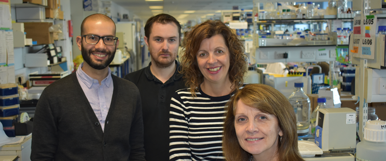
Dr Leandro Lemgruber Soares | Imaging TechnologistDr Lemgruber Soares is an Imaging Technologist who both established and manages the Glasgow Imaging Facility. Following time at the School of Infection & Immunuty, Leandro was appointed College Research Facility – Cellular Analysis Lead. |
 |
Ms Maragret Mullin | Electron Microscopy Technician |
 |
Mrs Susan Baillie | Research TechnicianUsing her knowledge of the many micrscopes we have on offer, Susan's role at the Glasgow Imaging Facility sees her train researchers in microscopy techniques. |
|
Mr Ryan Ritchie | IVIS Facility ManagerRyan Ritchie is the facility manager for the In Vivo Imaging System (IVIS), as well as currently working as one of Professor Mike Barrett’s laboratory technicians. |
 |

Guidelines
Access Charges to the Facility
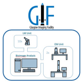
The Glasgow Imaging Facility (GIF) consists of the light microscopes located in the Sir Graeme Davies Building (SGDB) and the Electron Microscopy (EM) unit at the Joseph Black Building (JBB).
The full list of equipment available and their locations can be found below.
There is an annual charge for access to the light microscopes on top of the bench fees per person / financial year (from August to July), plus an hourly fee dependent on the equipment used.
If the maintenance contract of a specific equipment is covered by a grant, the members of that research group will not pay for the use of that microscope but will pay the annual access fee and the hourly charge of any other equipment used.
To help meet the cost of service contracts, all PIs must include appropriate hourly imaging costs in their grant proposals. The usage costs will be charged monthly through invoices to be sent directly to the PIs.
When writing a grant, the PIs should contact Dr Leandro Lemgruber for advice and guidance so that accurate costings are calculated for the applications. He can advise on the correct amount of time and the correct equipment to include in the proposal.
Glasgow Imaging Facility Publication Policy
As an academic core facility, GIF in common with other universities follows the internationally established guidelines in different universities and institutions, stipulated by journals and the publication houses.
We have, therefore, implemented a new publication policy within the facility:
- All publications resulting from the use of instruments within the facility should acknowledge GIF as a whole, e.g. 'The authors gratefully, acknowledge the Glasgow Imaging Facility for their support and assurance in this work'
- Where users have had significant help from a particular member of GIF staff or GIF staff have generated additional data personally, this staff member should be acknowledged by name, alongside the facility if applicable, e.g. 'The authors thank [insert name] of the Glasgow Imgaing Facility for their support and assistance in this work.
- If scientists from GIF contributed more than just routine techniques (for example, development or adaptation of protocols to suit samples or materials, designing or redesigning experiments, extensive data analysis and interpretation), it constitutes contributions meriting co-authorship in papers that use data.
We appreciate that authorship decisions are generally made at the preparation stage of a manuscript, rather than at the initiation of the work; GIF expect to be informed of any potential publications resulting from the work.
GIF staff should have the opportunity to participate in drafting the pertinent part of the paper and give final approval to the wording and conclusions drawn before publication, as any other contributing scientist would.
If you are uncertain about co-outhonhip or have any questions or concerns about this, please discuss this issue with the facility manager in advance.
Please send a reprint of the paper, or an email including the reference information for the publication, to the Glasgow Imaging Facility.
We also politely request that you remember to acknowledge use of GIF in your presentations and cite the facility in grants, as appropriate.
Imaging Techniques
The Institute and Research Centres have imaging technologies that cover whole body imaging (resolution in mm), through conventional wide-field, confocal and multi-photon microscopy (resolution in µm), to high-resolution approaches breaking the resolution limit of light (resolution in nm).
The microscopy technologies that are part of GIF are: Spinning Disk Confocal, Multiphoton, High Content InCell (previously part of the Scottish Bioscreening Facility), Super-Resolution (3D-SIM and STORM), wide-field deconvolution (DV-Core and Leica DMi8) and the imaging flow cytometer ImageStream.
In addition, the In vivo Imaging Systems (IVIS and FxPro) located in JRF and CRF respectively, plus the Electron Microscopy Unit, previously part of the School of Life Sciences, will be also integrated within 3IP. An additional system will be integrated later this year – a new upright Zeiss 880 confocal.


Showcase Slideshow
Bookings
To use any of the light microscopes in the Glasgow Imaging Facility, the user must be trained and the equipment booked before use.
Click the button below to book your slot.

Remember to book enough time for your experiment and saving the files.
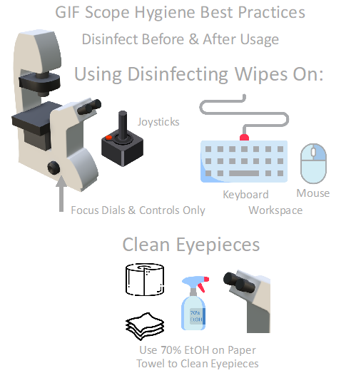
Microscope Cleaning