Brachial Plexus - Past and Present and Future
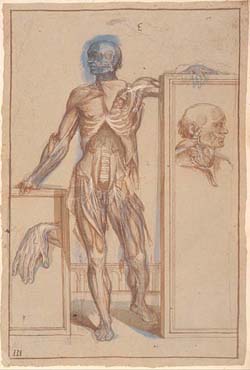
 As a trainee surgeon I form part of the team that looks after patients with damaged nerves and operate to repair them. An in-depth knowledge of nerve anatomy is necessary and is gleaned from the study of detailed anatomical dissections, such as those found in The Hunterian’s Anatomy collection.
I hope to explore the Brachial Plexus through the eyes of William Hunter, using items from his book collection in Special Collections
Microsurgery, with delicate instruments, an operative microscope and stitches finer than a hair, is used to repair injured nerves, however, despite careful technique not all of the injured nerve grows back. This leaves patients with numbness, pain and loss of movement. The brachial plexus is a large network of nerves supplying the upper limb and the Scottish National Brachial Plexus Centre is based in Glasgow (http://www.brachialplexus.scot.nhs.uk). Damage to brachial plexus can be functionally devastating and result in patients being unable to move their arm, with a clear impact on work, sport and play. New ways of addressing nerve injury at the cell-scale are needed to restore function after nerve injury. In my research I work as part of a team of scientists and surgeons developing bioengineering approaches to develop a novel approach to nerve repair and improve healing. This laboratory based project incorporates biodegradable polymer fabrication, 3-D printing and stem cell engineering to assist nerves to grow across the site of injury. It is necessary to confirm that these techniques are safe in the laboratory, before translation to clinical practice.
 Through this project I seek to explore the topic from another perspective and receive feedback, whilst introducing interested audiences to:
- the beautiful and complex anatomy of their own nervous system through interaction with Hunter’s nerve drawings and dissection specimens
- current nerve surgery – intraoperative descriptions and images and objects (e.g. safe display microsuture)
- bioengineering – properties of a good clinical material.
- 3-D printing on a hand-held device
- visualising fixed, stained stem cells.
Suzanne Thomson – 1st year PhD, Molecular Cell and Systems Biology, Centre for Cell Engineering

